7
views
0
recommends
+1 Recommend
1 collections
    0
    shares
      • Record: found
      • Abstract: found
      • Article: found
      Is Open Access

      Basophils beyond allergic and parasitic diseases

      review-article

      Read this article at

      Bookmark
          There is no author summary for this article yet. Authors can add summaries to their articles on ScienceOpen to make them more accessible to a non-specialist audience.

          Abstract

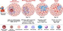
          Basophils bind IgE via FcεRI-αβγ 2, which they uniquely share only with mast cells. In doing so, they can rapidly release mediators that are hallmark of allergic disease. This fundamental similarity, along with some morphological features shared by the two cell types, has long brought into question the biological significance that basophils mediate beyond that of mast cells. Unlike mast cells, which mature and reside in tissues, basophils are released into circulation from the bone marrow (constituting 1% of leukocytes), only to infiltrate tissues under specific inflammatory conditions. Evidence is emerging that basophils mediate non-redundant roles in allergic disease and, unsuspectingly, are implicated in a variety of other pathologies [e.g., myocardial infarction, autoimmunity, chronic obstructive pulmonary disease, fibrosis, cancer, etc.]. Recent findings strengthen the notion that these cells mediate protection from parasitic infections, whereas related studies implicate basophils promoting wound healing. Central to these functions is the substantial evidence that human and mouse basophils are increasingly implicated as important sources of IL-4 and IL-13. Nonetheless, much remains unclear regarding the role of basophils in pathology vs. homeostasis. In this review, we discuss the dichotomous (protective and/or harmful) roles of basophils in a wide spectrum of non-allergic disorders.

          Related collections

          Most cited references277

          • Record: found
          • Abstract: not found
          • Article: not found

          Severe Covid-19

            Bookmark
            • Record: found
            • Abstract: found
            • Article: not found

            Polarization of tumor-associated neutrophil phenotype by TGF-beta: "N1" versus "N2" TAN.

            TGF-beta blockade significantly slows tumor growth through many mechanisms, including activation of CD8(+) T cells and macrophages. Here, we show that TGF-beta blockade also increases neutrophil-attracting chemokines, resulting in an influx of CD11b(+)/Ly6G(+) tumor-associated neutrophils (TANs) that are hypersegmented, more cytotoxic to tumor cells, and express higher levels of proinflammatory cytokines. Accordingly, following TGF-beta blockade, depletion of these neutrophils significantly blunts antitumor effects of treatment and reduces CD8(+) T cell activation. In contrast, in control tumors, neutrophil depletion decreases tumor growth and results in more activated CD8(+) T cells intratumorally. Together, these data suggest that TGF-beta within the tumor microenvironment induces a population of TAN with a protumor phenotype. TGF-beta blockade results in the recruitment and activation of TANs with an antitumor phenotype.
              Bookmark
              • Record: found
              • Abstract: found
              • Article: not found

              Cellular and molecular mechanisms of fibrosis.

              T A Wynn (2008)
              Fibrosis is defined by the overgrowth, hardening, and/or scarring of various tissues and is attributed to excess deposition of extracellular matrix components including collagen. Fibrosis is the end result of chronic inflammatory reactions induced by a variety of stimuli including persistent infections, autoimmune reactions, allergic responses, chemical insults, radiation, and tissue injury. Although current treatments for fibrotic diseases such as idiopathic pulmonary fibrosis, liver cirrhosis, systemic sclerosis, progressive kidney disease, and cardiovascular fibrosis typically target the inflammatory response, there is accumulating evidence that the mechanisms driving fibrogenesis are distinct from those regulating inflammation. In fact, some studies have suggested that ongoing inflammation is needed to reverse established and progressive fibrosis. The key cellular mediator of fibrosis is the myofibroblast, which when activated serves as the primary collagen-producing cell. Myofibroblasts are generated from a variety of sources including resident mesenchymal cells, epithelial and endothelial cells in processes termed epithelial/endothelial-mesenchymal (EMT/EndMT) transition, as well as from circulating fibroblast-like cells called fibrocytes that are derived from bone-marrow stem cells. Myofibroblasts are activated by a variety of mechanisms, including paracrine signals derived from lymphocytes and macrophages, autocrine factors secreted by myofibroblasts, and pathogen-associated molecular patterns (PAMPS) produced by pathogenic organisms that interact with pattern recognition receptors (i.e. TLRs) on fibroblasts. Cytokines (IL-13, IL-21, TGF-beta1), chemokines (MCP-1, MIP-1beta), angiogenic factors (VEGF), growth factors (PDGF), peroxisome proliferator-activated receptors (PPARs), acute phase proteins (SAP), caspases, and components of the renin-angiotensin-aldosterone system (ANG II) have been identified as important regulators of fibrosis and are being investigated as potential targets of antifibrotic drugs. This review explores our current understanding of the cellular and molecular mechanisms of fibrogenesis. 2007 Pathological Society of Great Britain and Ireland
                Bookmark

                Author and article information

                Contributors
                Journal
                Front Immunol
                Front Immunol
                Front. Immunol.
                Frontiers in Immunology
                Frontiers Media S.A.
                1664-3224
                02 May 2023
                2023
                02 May 2023
                : 14
                : 1190034
                Affiliations
                [1] 1 Department of Translational Medical Sciences, University of Naples Federico II , Naples, Italy
                [2] 2 World Allergy Organization (WAO), Center of Excellence (CoE) , Naples, Italy
                [3] 3 Center for Basic and Clinical Immunology Research (CISI), University of Naples Federico II , Naples, Italy
                [4] 4 Institute of Experimental Endocrinology and Oncology “G. Salvatore”, National Research Council (CNR) , Naples, Italy
                [5] 5 Division of Allergy and Clinical Immunology, The Johns Hopkins University School of Medicine , Baltimore, MD, United States
                Author notes

                Edited by: Christophe Pellefigues, CNRS EMR8252 Centre de Recherche sur l’Inflammation, France

                Reviewed by: Jagadeesh Bayry, Indian Institute of Technology Palakkad, India; Bernhard F. Gibbs, University of Oldenburg, Germany

                *Correspondence: Gilda Varricchi, gildanet@ 123456gmail.com ; John T. Schroeder, schray@ 123456jhmi.edu

                †ORCID: Remo Poto, orcid.org/0000-0002-4723-0167; Stefania Loffredo, orcid.org/0000-0002-5871-1898; Gianni Marone, orcid.org/0000-0002-9849-4701; Antonio Di Salvatore, orcid.org/0000-0002-6434-3112; Amato de Paulis, orcid.org/0000-0003-0347-2540; John T. Schroeder, orcid.org/0000-0002-5263-7850; Gilda Varricchi, orcid.org/0000-0002-9285-4657

                Article
                10.3389/fimmu.2023.1190034
                10185837
                37205111
                1bd5d3f2-61cb-4574-917a-8f3a616ee83a
                Copyright © 2023 Poto, Loffredo, Marone, Di Salvatore, de Paulis, Schroeder and Varricchi

                This is an open-access article distributed under the terms of the Creative Commons Attribution License (CC BY). The use, distribution or reproduction in other forums is permitted, provided the original author(s) and the copyright owner(s) are credited and that the original publication in this journal is cited, in accordance with accepted academic practice. No use, distribution or reproduction is permitted which does not comply with these terms.

                History
                : 20 March 2023
                : 14 April 2023
                Page count
                Figures: 6, Tables: 1, Equations: 0, References: 277, Pages: 19, Words: 8939
                Funding
                This work was supported in part by grants from the CISI-Lab Project (University of Naples Federico II), TIMING and Campania Bioscience Projects (Regione Campania) (GM and GV), and NIAID and NIH grants AI115703 and AI141486 (JS).
                Categories
                Immunology
                Review
                Custom metadata
                Molecular Innate Immunity

                Immunology
                alarmins,allergy,autoimmunity,basophil,cancer,covid-19, myocardial infarction
                Immunology
                alarmins, allergy, autoimmunity, basophil, cancer, covid-19, myocardial infarction

                Comments

                Comment on this article