118
views
0
recommends
+1 Recommend
0 collections
    0
    shares
      • Record: found
      • Abstract: found
      • Article: not found

      Autophagic degradation of farnesylated prelamin A as a therapeutic approach to lamin-linked progeria

      review-article

      Read this article at

      ScienceOpenPublisherPMC
      Bookmark
          There is no author summary for this article yet. Authors can add summaries to their articles on ScienceOpen to make them more accessible to a non-specialist audience.

          Abstract

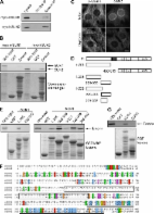
          Farnesylated prelamin A is a processing intermediate produced in the lamin A maturation pathway. Accumulation of a truncated farnesylated prelamin A form, called progerin, is a hallmark of the severe premature ageing syndrome, Hutchinson-Gilford progeria. Progerin elicits toxic effects in cells, leading to chromatin damage and cellular senescence and ultimately causes skin and endothelial defects, bone resorption, lipodystrophy and accelerated ageing. Knowledge of the mechanism underlying prelamin A turnover is critical for the development of clinically effective protein inhibitors that can avoid accumulation to toxic levels without impairing lamin A/C expression, which is essential for normal biological functions. Little is known about specific molecules that may target farnesylated prelamin A to elicit protein degradation. Here, we report the discovery of rapamycin as a novel inhibitor of progerin, which dramatically and selectively decreases protein levels through a mechanism involving autophagic degradation. Rapamycin treatment of progeria cells lowers progerin, as well as wild-type prelamin A levels, and rescues the chromatin phenotype of cultured fibroblasts, including histone methylation status and BAF and LAP2α distribution patterns. Importantly, rapamycin treatment does not affect lamin C protein levels, but increases the relative expression of the prelamin A endoprotease ZMPSTE24. Thus, rapamycin, an antibiotic belonging to the class of macrolides, previously found to increase longevity in mouse models, can serve as a therapeutic tool, to eliminate progerin, avoid farnesylated prelamin A accumulation, and restore chromatin dynamics in progeroid laminopathies.

          Related collections

          Most cited references17

          • Record: found
          • Abstract: found
          • Article: not found

          Autophagy is defective in collagen VI muscular dystrophies, and its reactivation rescues myofiber degeneration.

          Autophagy is crucial in the turnover of cell components, and clearance of damaged organelles by the autophagic-lysosomal pathway is essential for tissue homeostasis. Defects of this degradative system have a role in various diseases, but little is known about autophagy in muscular dystrophies. We have previously found that muscular dystrophies linked to collagen VI deficiency show dysfunctional mitochondria and spontaneous apoptosis, leading to myofiber degeneration. Here we demonstrate that this persistence of abnormal organelles and apoptosis are caused by defective autophagy. Skeletal muscles of collagen VI-knockout (Col6a1(-/-)) mice had impaired autophagic flux, which matched the lower induction of beclin-1 and BCL-2/adenovirus E1B-interacting protein-3 (Bnip3) and the lack of autophagosomes after starvation. Forced activation of autophagy by genetic, dietary and pharmacological approaches restored myofiber survival and ameliorated the dystrophic phenotype of Col6a1(-/-) mice. Furthermore, muscle biopsies from subjects with Bethlem myopathy or Ullrich congenital muscular dystrophy had reduced protein amounts of beclin-1 and Bnip3. These findings indicate that defective activation of the autophagic machinery is pathogenic in some congenital muscular dystrophies.
            Bookmark
            • Record: found
            • Abstract: found
            • Article: not found

            Combined treatment with statins and aminobisphosphonates extends longevity in a mouse model of human premature aging.

            Several human progerias, including Hutchinson-Gilford progeria syndrome (HGPS), are caused by the accumulation at the nuclear envelope of farnesylated forms of truncated prelamin A, a protein that is also altered during normal aging. Previous studies in cells from individuals with HGPS have shown that farnesyltransferase inhibitors (FTIs) improve nuclear abnormalities associated with prelamin A accumulation, suggesting that these compounds could represent a therapeutic approach for this devastating progeroid syndrome. We show herein that both prelamin A and its truncated form progerin/LADelta50 undergo alternative prenylation by geranylgeranyltransferase in the setting of farnesyltransferase inhibition, which could explain the low efficiency of FTIs in ameliorating the phenotypes of progeroid mouse models. We also show that a combination of statins and aminobisphosphonates efficiently inhibits both farnesylation and geranylgeranylation of progerin and prelamin A and markedly improves the aging-like phenotypes of mice deficient in the metalloproteinase Zmpste24, including growth retardation, loss of weight, lipodystrophy, hair loss and bone defects. Likewise, the longevity of these mice is substantially extended. These findings open a new therapeutic approach for human progeroid syndromes associated with nuclear-envelope abnormalities.
              Bookmark
              • Record: found
              • Abstract: found
              • Article: found
              Is Open Access

              Mammalian SUN Protein Interaction Networks at the Inner Nuclear Membrane and Their Role in Laminopathy Disease Processes*

              The nuclear envelope (NE) LINC complex, in mammals comprised of SUN domain and nesprin proteins, provides a direct connection between the nuclear lamina and the cytoskeleton, which contributes to nuclear positioning and cellular rigidity. SUN1 and SUN2 interact with lamin A, but lamin A is only required for NE localization of SUN2, and it remains unclear how SUN1 is anchored. Here, we identify emerin and short nesprin-2 isoforms as novel nucleoplasmic binding partners of SUN1/2. These have overlapping binding sites distinct from the lamin A binding site. However, we demonstrate that tight association of SUN1 with the nuclear lamina depends upon a short motif within residues 209–228, a region that does not interact significantly with known SUN1 binding partners. Moreover, SUN1 localizes correctly in cells lacking emerin. Importantly then, the major determinant of SUN1 NE localization has yet to be identified. We further find that a subset of lamin A mutations, associated with laminopathies Emery-Dreifuss muscular dystrophy (EDMD) and Hutchinson-Gilford progeria syndrome (HGPS), disrupt lamin A interaction with SUN1 and SUN2. Despite this, NE localization of SUN1 and SUN2 is not impaired in cell lines from either class of patients. Intriguingly, SUN1 expression at the NE is instead enhanced in a significant proportion of HGPS but not EDMD cells and strongly correlates with pre-lamin A accumulation due to preferential interaction of SUN1 with pre-lamin A. We propose that these different perturbations in lamin A-SUN protein interactions may underlie the opposing effects of EDMD and HGPS mutations on nuclear and cellular mechanics.
                Bookmark

                Author and article information

                Journal
                Eur J Histochem
                EJH
                EJH
                European Journal of Histochemistry : EJH
                PAGEPress Publications (Pavia, Italy )
                1121-760X
                2038-8306
                19 October 2011
                19 October 2011
                : 55
                : 4
                : e36
                Affiliations
                [1 ]National Research Council of Italy, Institute of Molecular Genetics, IGM-CNR, Unit of Bologna IOR, Bologna;
                [2 ]Laboratory of Musculoskeletal Cell Biology RAMSES, Rizzoli Orthopedic Institute, Bologna;
                [3 ]Department of Biopathology and Diagnostic Imaging Tor Vergata University, Rome;
                [4 ]National Agency for the Evaluation of Universities and Research, ANVUR, Rome Italy and San Pietro Fatebenefratelli Hospital, Rome;
                [5 ]Laboratory of Preclinical and Surgical Studies, Rizzoli Orthopedic Institute and BITTA, RIT, IOR, Bologna;
                [6 ]Department of Histology, University of Modena and Reggio Emilia, Modena;
                [7 ]Department of Pediatrics, S. Orsola-Malpighi Hospital, University of Bologna, Italy
                Author notes
                Correspondence: Dr. Giovanna Lattanzi, IGM-CNR, Unit of Bologna, via di Barbiano 1/10, 40136 Bologna, Italy. Tel. +39.051.6366394 - Fax: +39.051.583593. E-mail: giovanna.lattanzi@ 123456cnr.it
                Article
                ejh.2011.e36
                10.4081/ejh.2011.e36
                3284238
                22297442
                1b7a4f54-0e04-4f61-8375-987fd5b8aa0b
                ©Copyright V. Cenni et al., 2011

                This work is licensed under a Creative Commons Attribution NonCommercial 3.0 License (CC BY-NC 3.0).

                Licensee PAGEPress, Italy

                History
                : 12 July 2011
                : 10 August 2011
                Categories
                Original Paper

                Clinical chemistry
                rapamycin.,progeria,protein degradation,laminopathies,prelamin a,autophagy
                Clinical chemistry
                rapamycin., progeria, protein degradation, laminopathies, prelamin a, autophagy

                Comments

                Comment on this article