7
views
0
recommends
+1 Recommend
0 collections
    0
    shares
      • Record: found
      • Abstract: found
      • Article: found
      Is Open Access

      Catalpol promotes the osteogenic differentiation of bone marrow mesenchymal stem cells via the Wnt/β-catenin pathway

      research-article

      Read this article at

      Bookmark
          There is no author summary for this article yet. Authors can add summaries to their articles on ScienceOpen to make them more accessible to a non-specialist audience.

          Abstract

          Background

          Rehmanniae Radix is a traditional herbal medicine in East Asia that has been widely used to treat patients with osteoporosis. However, the effect of catalpol, the primary active principle component of Rehmanniae Radix, on the function of bone marrow mesenchymal stem cells (BMSCs) and the underlying molecular mechanisms associated with its activity remain poorly understood.

          Methods

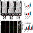
          The effect of catalpol on the proliferation of BMSCs was evaluated using a Cell Counting Kit-8 assay. Alkaline phosphatase (ALP) staining, ALP activity and Alizarin Red staining were performed to elucidate the effect of catalpol on the osteogenesis of BMSCs. qRT-PCR, Western blotting and immunofluorescence were performed to evaluate the expression of osteo-specific markers and the Wnt/β-catenin signalling-related genes and proteins. Moreover, a rat critical-sized calvarial defect model and a rat ovariectomy model were used to assess the effect of catalpol on bone regeneration in vivo.

          Results

          Catalpol significantly enhanced osteoblast-specific gene expression, alkaline phosphatase activity and calcium deposition in BMSCs in vitro. This phenomenon was accompanied by an upregulation of Wnt/β-catenin signalling. In addition, the enhanced osteogenesis due to catalpol treatment was partially reversed by a Wnt/β-catenin antagonist. Furthermore, catalpol increased the bone healing capacity of BMSCs in a rat critical-sized calvarial defect model and attenuated bone loss in a rat ovariectomy model.

          Conclusions

          These data suggest that catalpol enhances the osteogenic differentiation of BMSCs, partly via activation of the Wnt/β-catenin pathway. Catalpol may provide a new strategy for bone tissue engineering and can be a potential agent for the treatment of postmenopausal osteoporosis.

          Electronic supplementary material

          The online version of this article (10.1186/s13287-019-1143-y) contains supplementary material, which is available to authorized users.

          Related collections

          Most cited references37

          • Record: found
          • Abstract: found
          • Article: not found

          Regulation of bone mass by Wnt signaling.

          Wnt proteins are a family of secreted proteins that regulate many aspects of cell growth, differentiation, function, and death. Considerable progress has been made in our understanding of the molecular links between Wnt signaling and bone development and remodeling since initial reports that mutations in the Wnt coreceptor low-density lipoprotein receptor-related protein 5 (LRP5) are causally linked to alterations in human bone mass. Of the pathways activated by Wnts, it is signaling through the canonical (i.e., Wnt/beta-catenin) pathway that increases bone mass through a number of mechanisms including renewal of stem cells, stimulation of preosteoblast replication, induction of osteoblastogenesis, and inhibition of osteoblast and osteocyte apoptosis. This pathway is an enticing target for developing drugs to battle skeletal diseases as Wnt/beta-catenin signaling is composed of a series of molecular interactions that offer potential places for pharmacological intervention. In considering opportunities for anabolic drug discovery in this area, one must consider multiple factors, including (a) the roles of Wnt signaling for development, remodeling, and pathology of bone; (b) how pharmacological interventions that target this pathway may specifically treat osteoporosis and other aspects of skeletal health; and (c) whether the targets within this pathway are amenable to drug intervention. In this Review we discuss the current understanding of this pathway in terms of bone biology and assess whether targeting this pathway might yield novel therapeutics to treat typical bone disorders.
            Bookmark
            • Record: found
            • Abstract: found
            • Article: not found

            Severely suppressed bone turnover: a potential complication of alendronate therapy.

            Alendronate, an inhibitor of bone resorption, is widely used in osteoporosis treatment. However, concerns have been raised about potential oversuppression of bone turnover during long-term use. We report on nine patients who sustained spontaneous nonspinal fractures while on alendronate therapy, six of whom displayed either delayed or absent fracture healing for 3 months to 2 yr during therapy. Histomorphometric analysis of the cancellous bone showed markedly suppressed bone formation, with reduced or absent osteoblastic surface in most patients. Osteoclastic surface was low or low-normal in eight patients, and eroded surface was decreased in four. Matrix synthesis was markedly diminished, with absence of double-tetracycline label and absent or reduced single-tetracycline label in all patients. The same trend was seen in the intracortical and endocortical surfaces. Our findings raise the possibility that severe suppression of bone turnover may develop during long-term alendronate therapy, resulting in increased susceptibility to, and delayed healing of, nonspinal fractures. Although coadministration of estrogen or glucocorticoids appears to be a predisposing factor, this apparent complication can also occur with monotherapy. Our observations emphasize the need for increased awareness and monitoring for the potential development of excessive suppression of bone turnover during long-term alendronate therapy.
              Bookmark
              • Record: found
              • Abstract: found
              • Article: found
              Is Open Access

              Exosomes/tricalcium phosphate combination scaffolds can enhance bone regeneration by activating the PI3K/Akt signaling pathway

              Background Recently, accumulating evidence has shown that exosomes, the naturally secreted nanocarriers of cells, can exert therapeutic effects in various disease models in the absence of parent cells. However, application of exosomes in bone defect repair and regeneration has been rarely reported, and little is known regarding their underlying mechanisms. Methods Exosomes derived from human-induced pluripotent stem cell-derived mesenchymal stem cells (hiPS-MSC-Exos) were combined with tricalcium phosphate (β-TCP) to repair critical-sized calvarial bone defects, and the efficacy was assessed by histological examination. We evaluated the in vitro effects of hiPSC-MSC-Exos on the proliferation, migration, and osteogenic differentiation of human bone marrow-derived mesenchymal stem cells (hBMSCs) by cell-counting, scratch assays, and qRT-PCR, respectively. Gene expression profiling and bioinformatics analyses were also used to identify the underlying mechanisms in the repair. Results We found that the exosome/β-TCP combination scaffolds could enhance osteogenesis as compared to pure β-TCP scaffolds. In vitro assays showed that the exosomes could release from β-TCP and could be internalized by hBMSCs. In addition, the internalization of exosomes into hBMSCs could profoundly enhance the proliferation, migration, and osteogenic differentiation of hBMSCs. Furthermore, gene expression profiling and bioinformatics analyses demonstrated that exosome/β-TCP combination scaffolds significantly altered the expression of a network of genes involved in the PI3K/Akt signaling pathway. Functional studies further confirmed that the PI3K/Akt signaling pathway was the critical mediator during the exosome-induced osteogenic responses of hBMSCs. Conclusions We propose that the exosomes can enhance the osteoinductivity of β-TCP through activating the PI3K/Akt signaling pathway of hBMSCs, which means that the exosome/β-TCP combination scaffolds possess better osteogenesis activity than pure β-TCP scaffolds. These results indicate that naturally secreted nanocarriers-exosomes can be used as a bioactive material to improve the bioactivity of the biomaterials, and that hiPS-MSC-Exos combined with β-TCP scaffolds can be potentially used for repairing bone defects. Electronic supplementary material The online version of this article (doi:10.1186/s13287-016-0391-3) contains supplementary material, which is available to authorized users.
                Bookmark

                Author and article information

                Contributors
                06-zhuyu@sjtu.edu.cn
                ymwang6thhosp@163.com
                jiayachao0124@126.com
                +86 13611960282 , xujia0117@126.com
                +86 21 64369181 , ymchai@sjtu.edu.cn
                Journal
                Stem Cell Res Ther
                Stem Cell Res Ther
                Stem Cell Research & Therapy
                BioMed Central (London )
                1757-6512
                22 January 2019
                22 January 2019
                2019
                : 10
                : 37
                Affiliations
                ISNI 0000 0004 1798 5117, GRID grid.412528.8, Department of Orthopedic Surgery, , Shanghai Jiao Tong University Affiliated Sixth People’s Hospital, ; Yishan Rd 600, Shanghai, 200233 People’s Republic of China
                Author information
                http://orcid.org/0000-0002-1972-5971
                Article
                1143
                10.1186/s13287-019-1143-y
                6341609
                30670092
                1a8b52a0-be3e-4a90-a008-f2579d3f3178
                © The Author(s). 2019

                Open AccessThis article is distributed under the terms of the Creative Commons Attribution 4.0 International License ( http://creativecommons.org/licenses/by/4.0/), which permits unrestricted use, distribution, and reproduction in any medium, provided you give appropriate credit to the original author(s) and the source, provide a link to the Creative Commons license, and indicate if changes were made. The Creative Commons Public Domain Dedication waiver ( http://creativecommons.org/publicdomain/zero/1.0/) applies to the data made available in this article, unless otherwise stated.

                History
                : 13 August 2018
                : 3 January 2019
                : 9 January 2019
                Funding
                Funded by: FundRef http://dx.doi.org/10.13039/501100001809, National Natural Science Foundation of China;
                Award ID: 81772338
                Award ID: 81572122
                Funded by: Interdisciplinary Program of Shanghai Jiao Tong University
                Award ID: YG2017ZD05
                Funded by: Shanghai Leading Talent Plan
                Award ID: 043
                Categories
                Research
                Custom metadata
                © The Author(s) 2019

                Molecular medicine
                catalpol,bone marrow mesenchymal stem cells,wnt/β-catenin signalling,bone regeneration

                Comments

                Comment on this article