35
views
0
recommends
+1 Recommend
0 collections
    5
    shares
      • Record: found
      • Abstract: found
      • Article: found
      Is Open Access

      The small GTPases ARL-13 and ARL-3 coordinate intraflagellar transport and ciliogenesis

      research-article

      Read this article at

      Bookmark
          There is no author summary for this article yet. Authors can add summaries to their articles on ScienceOpen to make them more accessible to a non-specialist audience.

          Abstract

          Cilia intraflagellar transport and ciliogenesis are regulated by two small GTPases that maintain binding between IFT subcomplexes.

          Abstract

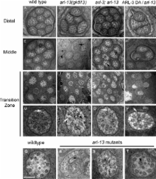
          Intraflagellar transport (IFT) machinery mediates the bidirectional movement of cargos that are required for the assembly and maintenance of cilia. However, little is known about how IFT is regulated in vivo. In this study, we show that the small guanosine triphosphatase (GTPase) adenosine diphosphate ribosylation factor–like protein 13 (ARL-13) encoded by the Caenorhabditis elegans homologue of the human Joubert syndrome causal gene ARL13B, localizes exclusively to the doublet segment of the cilium. arl-13 mutants have shortened cilia with various ultrastructural deformities and a disrupted association between IFT subcomplexes A and B. Intriguingly, depletion of ARL-3, another ciliary small GTPase, partially suppresses ciliogenesis defects in arl-13 mutants by indirectly restoring binding between IFT subcomplexes A and B. Rescue of arl-13 mutants by ARL-3 depletion is mediated by an HDAC6 deacetylase-dependent pathway. Thus, we propose that two conserved small GTPases, ARL-13 and ARL-3, coordinate to regulate IFT and that perturbing this balance results in cilia deformation.

          Related collections

          Most cited references44

          • Record: found
          • Abstract: found
          • Article: not found

          The ciliopathies: an emerging class of human genetic disorders.

          Cilia and flagella are ancient, evolutionarily conserved organelles that project from cell surfaces to perform diverse biological roles, including whole-cell locomotion; movement of fluid; chemo-, mechano-, and photosensation; and sexual reproduction. Consistent with their stringent evolutionary conservation, defects in cilia are associated with a range of human diseases, such as primary ciliary dyskinesia, hydrocephalus, polycystic liver and kidney disease, and some forms of retinal degeneration. Recent evidence indicates that ciliary defects can lead to a broader set of developmental and adult phenotypes, with mutations in ciliary proteins now associated with nephronophthisis, Bardet-Biedl syndrome, Alstrom syndrome, and Meckel-Gruber syndrome. The molecular data linking seemingly unrelated clinical entities are beginning to highlight a common theme, where defects in ciliary structure and function can lead to a predictable phenotypic pattern that has potentially predictive and therapeutic value.
            Bookmark
            • Record: found
            • Abstract: found
            • Article: not found

            Mutant sensory cilia in the nematode Caenorhabditis elegans.

            Eight classes of chemosensory neurons in C. elegans fill with fluorescein when living animals are placed in a dye solution. Fluorescein enters the neurons through their exposed sensory cilia. Mutations in 14 genes prevent dye uptake and disrupt chemosensory behaviors. Each of these genes affects the ultrastructure of the chemosensory cilia or their accessory cells. In each case, the cilia are shorter or less exposed than normal, suggesting that dye contact is the principal factor under selection. Ten genes affect many or all of the sensory cilia in the head. The daf-19 (m86) mutation eliminates all cilia, leaving only occasional centrioles in the dendrites. The cilia in che-13 (e1805), osm-1 (p808), osm-5 (p813), and osm-6 (p811) mutants have normal transition zones and severely shortened axonemes. Doublet-microtubules, attached to the membrane by Y links, assemble ectopically proximal to the cilia in these mutants. The amphid cilia in che-11 (e1810) are irregular in diameter and contain dark ground material in the middle of the axonemes. Certain mechanocilia are also affected. The amphid cilia in che-10 (e1809) apparently degenerate, leaving dendrites with bulb-shaped endings filled with dark ground material. The mechanocilia lack striated rootlets. Cilia defects have also been found in che-2, che-3, and daf-10 mutants. The osm-3 (p802) mutation specifically eliminates the distal segment of the amphid cilia. Mutations in three genes affect sensillar support cells. The che-12 (e1812) mutation eliminates matrix material normally secreted by the amphid sheath cell. The che-14 (e1960) mutation disrupts the joining of the amphid sheath and socket cells to form the receptor channel. A similar defect has been observed in daf-6 mutants. Four additional genes affect specific classes of ciliated sensory neurons. The mec-1 and mec-8 (e398) mutations disrupt the fasciculation of the amphid cilia. The cat-6 (e1861) mutation disrupts the tubular bodies of the CEP mechanocilia. A cryophilic thermotaxis mutant, ttx-1 (p767), lacks fingers on the AFD dendrite, suggesting this neuron is thermosensory.
              Bookmark
              • Record: found
              • Abstract: found
              • Article: not found

              HDAC6 modulates cell motility by altering the acetylation level of cortactin.

              Histone deacetylase 6 (HDAC6) is a tubulin-specific deacetylase that regulates microtubule-dependent cell movement. In this study, we identify the F-actin-binding protein cortactin as a HDAC6 substrate. We demonstrate that HDAC6 binds cortactin and that overexpression of HDAC6 leads to hypoacetylation of cortactin, whereas inhibition of HDAC6 activity leads to cortactin hyperacetylation. HDAC6 alters the ability of cortactin to bind F-actin by modulating a "charge patch" in its repeat region. Introduction of charge-preserving or charge-neutralizing mutations in this cortactin repeat region correlates with the gain or loss of F-actin binding ability, respectively. Cells expressing a charge-neutralizing cortactin mutant were less motile than control cells or cells expressing a charge-preserving mutant. These findings suggest that, in addition to its role in microtubule-dependent cell motility, HDAC6 influences actin-dependent cell motility by altering the acetylation status of cortactin, which, in turn, changes the F-actin binding activity of cortactin.
                Bookmark

                Author and article information

                Journal
                J Cell Biol
                J. Cell Biol
                jcb
                The Journal of Cell Biology
                The Rockefeller University Press
                0021-9525
                1540-8140
                14 June 2010
                : 189
                : 6
                : 1039-1051
                Affiliations
                [1 ]Division of Nephrology and Hypertension, Department of Internal Medicine , and [2 ]Department of Biochemistry and Molecular Biology, Mayo Clinic, Rochester, MN 55905
                Author notes
                Correspondence to Jinghua Hu: hu.jinghua@ 123456mayo.edu
                Article
                200912001
                10.1083/jcb.200912001
                2886347
                20530210
                180c183e-7759-4e06-8923-2b0f91692c1c
                © 2010 Li et al.

                This article is distributed under the terms of an Attribution–Noncommercial–Share Alike–No Mirror Sites license for the first six months after the publication date (see http://www.rupress.org/terms). After six months it is available under a Creative Commons License (Attribution–Noncommercial–Share Alike 3.0 Unported license, as described at http://creativecommons.org/licenses/by-nc-sa/3.0/).

                History
                : 1 December 2009
                : 13 May 2010
                Categories
                Research Articles
                Article

                Cell biology
                Cell biology

                Comments

                Comment on this article