19
views
0
recommends
+1 Recommend
0 collections
    0
    shares
      • Record: found
      • Abstract: found
      • Article: found
      Is Open Access

      Astragaloside IV protects against podocyte injury via SERCA2-dependent ER stress reduction and AMPKα-regulated autophagy induction in streptozotocin-induced diabetic nephropathy

      research-article

      Read this article at

      Bookmark
          There is no author summary for this article yet. Authors can add summaries to their articles on ScienceOpen to make them more accessible to a non-specialist audience.

          Abstract

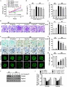
          Aberrant endoplasmic reticulum (ER) stress and autophagy are associated with diabetic nephropathy. Here we investigated the effect of astragaloside IV (AS-IV) on the progression of diabetic nephropathy (DN) and the underlying mechanism involving ER stress and autophagy in streptozotocin (STZ)-induced diabetic mice and high glucose (HG)-incubated podocytes. The diabetic mice developed progressive albuminuria and glomerulosclerosis within 8 weeks, which were significantly ameliorated by AS-IV treatment in a dose-dependent manner. Moreover, diabetes or HG-induced podocyte apoptosis was markedly attenuated by AS-IV, paralleled by a marked remission in ER stress and a remarkable restoration in impaired autophagy, which were associated with a significant improvement in the expression of sarcoendoplasmic reticulum Ca 2+ ATPase 2b (SERCA2b) and AMP-activated protein kinase α (AMPKα) phosphorylation, respectively. Knockdown of SERCA2 in podocytes induced ER stress and largely abolished the protective effect of AS-IV, but had no obvious effect on the expression of autophagy-associated proteins. On the other hand, blockade of either autophagy induction or AMPKα activation could also significantly mitigate AS-IV-induced beneficial effect. Collectively, these results suggest that AS-IV prevented the progression of DN, which is mediated at least in part by SERCA2-dependent ER stress attenuation and AMPKα-promoted autophagy induction.

          Related collections

          Most cited references32

          • Record: found
          • Abstract: found
          • Article: not found

          The role of inflammatory cytokines in diabetic nephropathy.

          Cytokines act as pleiotropic polypeptides regulating inflammatory and immune responses through actions on cells. They provide important signals in the pathophysiology of a range of diseases, including diabetes mellitus. Chronic low-grade inflammation and activation of the innate immune system are closely involved in the pathogenesis of diabetes and its microvascular complications. Inflammatory cytokines, mainly IL-1, IL-6, and IL-18, as well as TNF-alpha, are involved in the development and progression of diabetic nephropathy. In this context, cytokine genetics is of special interest to combinatorial polymorphisms among cytokine genes, their functional variations, and general susceptibility to diabetic nephropathy. Finally, the recognition of these molecules as significant pathogenic mediators in diabetic nephropathy leaves open the possibility of new potential therapeutic targets.
            Bookmark
            • Record: found
            • Abstract: found
            • Article: not found

            Rearrangements of the cytoskeleton and cell contacts induce process formation during differentiation of conditionally immortalized mouse podocyte cell lines.

            Mature podocytes are among the most complex differentiated cells and possess a highly branched array of foot processes that are essential to glomerular filtration in the kidney. Such differentiated podocytes are unable to replicate and culturing of primary podocytes results in rapid growth arrest. Therefore, conditionally immortalized mouse podocyte clones (MPC) were established, which are highly proliferative when cultured under permissive conditions. Nonpermissive conditions render the majority of MPC cells growth arrested within 6 days and induce many characteristics of differentiated podocytes. Both proliferating and differentiating MPC cells express the WT-1 protein and an ordered array of actin fibers and microtubules extends into the forming cellular processes during differentiation, reminiscent of podocyte processes in vivo. These cytoskeletal rearrangements and process formation are accompanied by the onset of synaptopodin synthesis, an actin-associated protein marking specifically differentiated podocytes. In addition, focal contacts are rearranged into an ordered pattern in differentiating MPC cells. Most importantly, electrophysiological studies demonstrate that differentiated MPC cells respond to the vasoactive peptide bradykinin by changes in intracellular calcium concentration. These results suggest a regulatory role of podocytes in glomerular filtration. Taken together, these studies establish that conditionally immortalized MPC cells retain a differentiation potential similar to podocytes in vivo. Therefore, the determinative steps of podocyte differentiation and process formation are studied for the first time using an inducible in vitro model.
              Bookmark
              • Record: found
              • Abstract: found
              • Article: not found

              Microalbuminuria and the risk for early progressive renal function decline in type 1 diabetes.

              This study aimed to establish the time of initiation and the determinants of renal function decline in type 1 diabetes. Until now, such decline has been assumed to be a late-occurring event associated with proteinuria. A total of 267 patients with normoalbuminuria and 301 patients with microalbuminuria were followed for 8 to 12 yr. Linear trends (slopes) in GFR were estimated by serial measurement of serum cystatin C. Cases of early renal function decline were defined by loss in cystatin C GFR that exceeded -3.3%/yr, a threshold that corresponds to the 2.5th percentile of the distribution of GFR slopes in an independent nondiabetic normotensive population. Cases of early renal function decline occurred in 9% (mean slope -4.4; range -5.9 to -3.3%/yr) of the normoalbuminuria group and 31% (mean slope -7.1; range -23.8 to -3.3%/yr) of the microalbuminuria group (P < 0.001). Risk for early renal function decline depended on whether microalbuminuria regressed, remained stable, or progressed, rising from 16 to 32 and 68%, respectively (P < 0.001). In multivariate analysis, risk for decline was higher after age 35 yr or when glycosylated hemoglobin exceeded 9% but did not vary with diabetes duration, smoking, BP, or angiotensin-converting enzyme inhibitor treatment. Contrary to the existing paradigm of diabetic nephropathy, progressive renal function decline in type 1 diabetes is an early event that occurs in a large proportion of patients with microalbuminuria. Together with testing for microalbuminuria, clinical protocols using cystatin C to diagnose early renal function decline and track response to therapeutic interventions should be developed.
                Bookmark

                Author and article information

                Contributors
                caoaili0312@sina.cn
                pengwen_01@vip.sina.com
                Journal
                Sci Rep
                Sci Rep
                Scientific Reports
                Nature Publishing Group UK (London )
                2045-2322
                31 July 2017
                31 July 2017
                2017
                : 7
                : 6852
                Affiliations
                [1 ]ISNI 0000 0001 2372 7462, GRID grid.412540.6, Laboratory of Renal Disease, , Putuo Hospital, Shanghai University of Traditional Chinese Medicine, ; Shanghai, 200062 China
                [2 ]ISNI 0000 0001 2372 7462, GRID grid.412540.6, Department of Nephrology, Putuo Hospital, , Shanghai University of Traditional Chinese Medicine, ; Shanghai, 200062 China
                [3 ]ISNI 0000 0001 0125 2443, GRID grid.8547.e, Department of Pharmacology, School of Pharmacy, , Fudan University, ; Shanghai, 201203 China
                Article
                7061
                10.1038/s41598-017-07061-7
                5537362
                28761152
                180322c4-c3a3-4d4b-9c75-768f29a8cc37
                © The Author(s) 2017

                Open Access This article is licensed under a Creative Commons Attribution 4.0 International License, which permits use, sharing, adaptation, distribution and reproduction in any medium or format, as long as you give appropriate credit to the original author(s) and the source, provide a link to the Creative Commons license, and indicate if changes were made. The images or other third party material in this article are included in the article’s Creative Commons license, unless indicated otherwise in a credit line to the material. If material is not included in the article’s Creative Commons license and your intended use is not permitted by statutory regulation or exceeds the permitted use, you will need to obtain permission directly from the copyright holder. To view a copy of this license, visit http://creativecommons.org/licenses/by/4.0/.

                History
                : 30 January 2017
                : 26 June 2017
                Categories
                Article
                Custom metadata
                © The Author(s) 2017

                Uncategorized
                Uncategorized

                Comments

                Comment on this article