4
views
0
recommends
+1 Recommend
0 collections
    0
    shares
      • Record: found
      • Abstract: found
      • Article: found
      Is Open Access

      Gallbladder Ciliated Foregut Cyst Suspected of Malignancy Preoperatively

      case-report

      Read this article at

      Bookmark
          There is no author summary for this article yet. Authors can add summaries to their articles on ScienceOpen to make them more accessible to a non-specialist audience.

          Abstract

          Background

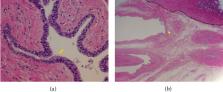
          Gallbladder ciliated foregut cysts (CFCs) of the lower diaphragm are extremely rare. Furthermore, they are rarely suspected of malignancy preoperatively. Case Presentation. A 50-year-old woman was referred to our hospital for further examination and treatment of a gallbladder tumor that was detected using abdominal ultrasonography (US). After a close inspection, she was diagnosed with a gallbladder tumor that was possibly malignant. Accordingly, open whole layer cholecystectomy was performed because intraoperative US revealed a tumor located on the intraperitoneal side of the gallbladder, and a rapid intraoperative pathological diagnosis identified no malignancy. A postoperative pathological examination revealed a cystic lesion with thin walls covered with ciliated epithelium, which laid on a connective tissue with smooth muscle fibers. Based on the above results, the final pathological diagnosis was CFC of the gallbladder without malignancy.

          Conclusions

          Cases of gallbladder CFC can be considered as cysts requiring treatment owing to CFCs' potential for malignant transformation and high-frequency symptoms.

          Related collections

          Most cited references22

          • Record: found
          • Abstract: not found
          • Article: not found

          Clinical practice guidelines for the management of biliary tract cancers 2019: The 3rd English edition

            Bookmark
            • Record: found
            • Abstract: found
            • Article: not found

            Squamous cell carcinoma arising in a ciliated hepatic foregut cyst.

            We report a case of squamous-cell carcinoma arising in a ciliated hepatic foregut cyst that occurred in a 21-year-old man. The cystic lesion was first discovered during childhood with no further follow-up. Following important weight loss over several months, the patient was admitted to our hospital where a CT scan showed a cystic and solid mass in segments V and VI of the liver involving the transverse mesocolon and the gastric antrum. A right hepatectomy with en-bloc right hemicolectomy and partial gastrectomy was performed. Gross examination showed a partially cystic liver mass with a maximum dimension of 10 cm infiltrating the large bowel wall. Microscopically, it was a poorly differentiated squamous-cell carcinoma arising from the wall of a liver cyst lined by a ciliated, pseudostratified columnar epithelium. Hepatic foregut cysts are uncommon, congenital, benign lesions that, when discovered, deserve careful clinical follow-up as malignant transformation, albeit exceptional, is possible.
              Bookmark
              • Record: found
              • Abstract: found
              • Article: not found

              Bronchogenic cyst in the abdomen.

              A bronchogenic cyst was found in the abdomen, in the retroperitoneum adjacent to the superior pancreatic body. The cyst was unilocular and contained about 100 ml pale yellow mucinous fluid. Microscopic examination revealed a pseudostratified columnar ciliated or cuboidal epithelium, seromucous glands, smooth muscle and cartilage, the distinctive features of bronchogenic cysts. This aberrant location of the cyst is explicable if abnormal buds of the tracheobronchial tree are pinched off and migrate into the abdomen in an early embryonic stage before the canal linking the abdominal with the thoracic cavity is closed by fusion of the future components of the diaphragm.
                Bookmark

                Author and article information

                Contributors
                Journal
                Case Rep Surg
                Case Rep Surg
                CRIS
                Case Reports in Surgery
                Hindawi
                2090-6900
                2090-6919
                2021
                21 October 2021
                : 2021
                : 6222947
                Affiliations
                1Department of Surgery, Kitakyushu Municipal Medical Center, Kitakyushu, Japan
                2Department of Pathology, Kitakyushu Municipal Medical Center, Kitakyushu, Japan
                Author notes

                Academic Editor: Tahsin Colak

                Author information
                https://orcid.org/0000-0003-0907-4666
                https://orcid.org/0000-0003-2148-4852
                Article
                10.1155/2021/6222947
                8553437
                16c954ae-0a06-4634-b043-8728bc53bdd4
                Copyright © 2021 Chikanori Tsutsumi et al.

                This is an open access article distributed under the Creative Commons Attribution License, which permits unrestricted use, distribution, and reproduction in any medium, provided the original work is properly cited.

                History
                : 30 July 2021
                : 18 September 2021
                Funding
                Funded by: Fukuoka Foundation for Sound Health
                Funded by: Shinnihon Foundation of Advanced Medical Treatment Research
                Funded by: Japan Society for the Promotion of Science
                Award ID: 19K23918
                Categories
                Case Report

                Surgery
                Surgery

                Comments

                Comment on this article