0
views
0
recommends
+1 Recommend
0 collections
    0
    shares
      • Record: found
      • Abstract: found
      • Article: found
      Is Open Access

      Functional ultrasound localization microscopy reveals brain-wide neurovascular activity on a microscopic scale

      research-article

      Read this article at

      Bookmark
          There is no author summary for this article yet. Authors can add summaries to their articles on ScienceOpen to make them more accessible to a non-specialist audience.

          Abstract

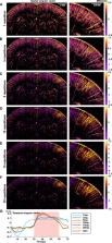
          The advent of neuroimaging has increased our understanding of brain function. While most brain-wide functional imaging modalities exploit neurovascular coupling to map brain activity at millimeter resolutions, the recording of functional responses at microscopic scale in mammals remains the privilege of invasive electrophysiological or optical approaches, but is mostly restricted to either the cortical surface or the vicinity of implanted sensors. Ultrasound localization microscopy (ULM) has achieved transcranial imaging of cerebrovascular flow, up to micrometre scales, by localizing intravenously injected microbubbles; however, the long acquisition time required to detect microbubbles within microscopic vessels has so far restricted ULM application mainly to microvasculature structural imaging. Here we show how ULM can be modified to quantify functional hyperemia dynamically during brain activation reaching a 6.5-µm spatial and 1-s temporal resolution in deep regions of the rat brain.

          Abstract

          Functional ultrasound localization microscopy monitors cerebrovascular blood flow by detecting the flow of injected microbubbles, providing access to brain activity at high spatiotemporal resolution.

          Related collections

          Most cited references58

          • Record: found
          • Abstract: found
          • Article: not found

          Imaging intracellular fluorescent proteins at nanometer resolution.

          We introduce a method for optically imaging intracellular proteins at nanometer spatial resolution. Numerous sparse subsets of photoactivatable fluorescent protein molecules were activated, localized (to approximately 2 to 25 nanometers), and then bleached. The aggregate position information from all subsets was then assembled into a superresolution image. We used this method--termed photoactivated localization microscopy--to image specific target proteins in thin sections of lysosomes and mitochondria; in fixed whole cells, we imaged vinculin at focal adhesions, actin within a lamellipodium, and the distribution of the retroviral protein Gag at the plasma membrane.
            Bookmark
            • Record: found
            • Abstract: found
            • Article: not found

            The Neurovascular Unit Coming of Age: A Journey through Neurovascular Coupling in Health and Disease

            The concept of neurovascular unit (NVU), formalized at the 2001 Stroke Progress Review Group meeting of the National Institute of Neurological Disorders and Stroke, emphasizes the intimate relationship between the brain and its vessels. Since then, the NVU has attracted the interest of the neuroscience community resulting in considerable advances in the field. Here the current state-of-knowledge of the NVU will be assessed, focusing on one of its most vital roles: the coupling between neural activity and blood flow. The evidence supports a conceptual shift in the mechanisms of neurovascular coupling, from a unidimensional process involving neuronal-astrocytic signaling to local blood vessels, to a multidimensional one in which mediators released from multiple cells engage distinct signaling pathways and effector systems across the entire cerebrovascular network in a highly orchestrated manner. The recently appreciated NVU dysfunction in neurodegenerative diseases, although still poorly understood, supports emerging concepts that maintaining neurovascular health promotes brain health.
              Bookmark
              • Record: found
              • Abstract: found
              • Article: not found

              Neurophysiological investigation of the basis of the fMRI signal.

              Functional magnetic resonance imaging (fMRI) is widely used to study the operational organization of the human brain, but the exact relationship between the measured fMRI signal and the underlying neural activity is unclear. Here we present simultaneous intracortical recordings of neural signals and fMRI responses. We compared local field potentials (LFPs), single- and multi-unit spiking activity with highly spatio-temporally resolved blood-oxygen-level-dependent (BOLD) fMRI responses from the visual cortex of monkeys. The largest magnitude changes were observed in LFPs, which at recording sites characterized by transient responses were the only signal that significantly correlated with the haemodynamic response. Linear systems analysis on a trial-by-trial basis showed that the impulse response of the neurovascular system is both animal- and site-specific, and that LFPs yield a better estimate of BOLD responses than the multi-unit responses. These findings suggest that the BOLD contrast mechanism reflects the input and intracortical processing of a given area rather than its spiking output.
                Bookmark

                Author and article information

                Contributors
                Mickael.tanter@espci.fr
                Journal
                Nat Methods
                Nat Methods
                Nature Methods
                Nature Publishing Group US (New York )
                1548-7091
                1548-7105
                4 August 2022
                4 August 2022
                2022
                : 19
                : 8
                : 1004-1012
                Affiliations
                GRID grid.440907.e, ISNI 0000 0004 1784 3645, Institute Physics for Medicine Paris, , INSERM U1273, ESPCI PSL Paris, CNRS UMR 8631, PSL Research University, ; Paris, France
                Author information
                http://orcid.org/0000-0002-3305-3315
                http://orcid.org/0000-0001-7739-8051
                Article
                1549
                10.1038/s41592-022-01549-5
                9352591
                35927475
                162e1a11-cb5f-442f-b67c-be5f4477d8b3
                © The Author(s) 2022

                Open Access This article is licensed under a Creative Commons Attribution 4.0 International License, which permits use, sharing, adaptation, distribution and reproduction in any medium or format, as long as you give appropriate credit to the original author(s) and the source, provide a link to the Creative Commons license, and indicate if changes were made. The images or other third party material in this article are included in the article’s Creative Commons license, unless indicated otherwise in a credit line to the material. If material is not included in the article’s Creative Commons license and your intended use is not permitted by statutory regulation or exceeds the permitted use, you will need to obtain permission directly from the copyright holder. To view a copy of this license, visit http://creativecommons.org/licenses/by/4.0/.

                History
                : 1 September 2021
                : 14 June 2022
                Funding
                Funded by: FundRef https://doi.org/10.13039/501100001961, AXA Research Fund (Le Fonds AXA pour la Recherche);
                Award ID: New Hopes in Medical Imaging with Ultrasound
                Award ID: New Hopes in Medical Imaging with Ultrasound
                Award ID: New Hopes in Medical Imaging with Ultrasound
                Award ID: New Hopes in Medical Imaging with Ultrasound
                Award ID: New Hopes in Medical Imaging with Ultrasound
                Award ID: New Hopes in Medical Imaging with Ultrasound
                Award Recipient :
                Categories
                Article
                Custom metadata
                © The Author(s), under exclusive licence to Springer Nature America, Inc. 2022

                Life sciences
                ultrasound,preclinical research,rat,sensorimotor processing
                Life sciences
                ultrasound, preclinical research, rat, sensorimotor processing

                Comments

                Comment on this article