120
views
0
recommends
+1 Recommend
2 collections
    0
    shares
      • Record: found
      • Abstract: found
      • Article: found
      Is Open Access

      Characterization of spike glycoprotein of SARS-CoV-2 on virus entry and its immune cross-reactivity with SARS-CoV

      research-article

      Read this article at

      Bookmark
          There is no author summary for this article yet. Authors can add summaries to their articles on ScienceOpen to make them more accessible to a non-specialist audience.

          Abstract

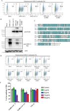
          Since 2002, beta coronaviruses (CoV) have caused three zoonotic outbreaks, SARS-CoV in 2002–2003, MERS-CoV in 2012, and the newly emerged SARS-CoV-2 in late 2019. However, little is currently known about the biology of SARS-CoV-2. Here, using SARS-CoV-2 S protein pseudovirus system, we confirm that human angiotensin converting enzyme 2 (hACE2) is the receptor for SARS-CoV-2, find that SARS-CoV-2 enters 293/hACE2 cells mainly through endocytosis, that PIKfyve, TPC2, and cathepsin L are critical for entry, and that SARS-CoV-2 S protein is less stable than SARS-CoV S. Polyclonal anti-SARS S1 antibodies T62 inhibit entry of SARS-CoV S but not SARS-CoV-2 S pseudovirions. Further studies using recovered SARS and COVID-19 patients’ sera show limited cross-neutralization, suggesting that recovery from one infection might not protect against the other. Our results present potential targets for development of drugs and vaccines for SARS-CoV-2.

          Abstract

          SARS-CoV-2 has spread globally. Here, the authors characterize the entry pathway of SARS-CoV-2, show that the SARS-CoV-2 spike protein is less stable than that of SARS-CoV, and show limited cross-neutralization activities between SARS-CoV and SARS-CoV-2 sera.

          Related collections

          Most cited references12

          • Record: found
          • Abstract: found
          • Article: not found

          Ebola virus. Two-pore channels control Ebola virus host cell entry and are drug targets for disease treatment.

          Ebola virus causes sporadic outbreaks of lethal hemorrhagic fever in humans, but there is no currently approved therapy. Cells take up Ebola virus by macropinocytosis, followed by trafficking through endosomal vesicles. However, few factors controlling endosomal virus movement are known. Here we find that Ebola virus entry into host cells requires the endosomal calcium channels called two-pore channels (TPCs). Disrupting TPC function by gene knockout, small interfering RNAs, or small-molecule inhibitors halted virus trafficking and prevented infection. Tetrandrine, the most potent small molecule that we tested, inhibited infection of human macrophages, the primary target of Ebola virus in vivo, and also showed therapeutic efficacy in mice. Therefore, TPC proteins play a key role in Ebola virus infection and may be effective targets for antiviral therapy.
            Bookmark
            • Record: found
            • Abstract: found
            • Article: not found

            Localization of neutralizing epitopes and the receptor-binding site within the amino-terminal 330 amino acids of the murine coronavirus spike protein.

            To localize the epitopes recognized by monoclonal antibodies (MAbs) specific for the S1 subunit of the murine coronavirus JHMV spike protein, we have expressed S1 proteins with different deletions from the C terminus of S1. S1utt is composed of the entire 769-amino-acid (aa) S1 protein; S1NM, S1N, S1n(330), and S1n(220) are deletion mutants with 594, 453, 330, and 220 aa from the N terminus of the S1 protein. The expressed S1 deletion mutant proteins were examined for reactivities to a panel of MAbs. All MAbs classified in groups A and B, those reactive to most mouse hepatitis virus (MHV) strains and those specific for isolate JHMV, respectively, recognized S1N(330) and the larger S1 deletion mutants but failed to react with S1N(220). MAbs in group C, specific for the larger S protein of JHMV, reacted only with the S1utt protein without any deletion. These results indicated that the domain composed of the N-terminal 330 aa comprised the cluster of conformational epitopes recognized by MAbs in groups A and B. It was also shown that the epitopes of MAbs in group C were not restricted to the region missing in the smaller S protein. These results together with the fact that all MAbs in group B retained high neutralizing activity suggested the possibility that the N-terminal 330 aa are responsible for binding to the MHV-specific receptors. In investigate this possibility, we expressed the receptor protein and examined the binding of each S1 deletion mutant to the receptor. It was demonstrated that the S1N(330) protein as well as other S1 deletion mutants larger than S1N(330) bound to the receptor. These results indicated that a domain composed of 330 aa at the N terminus of the S1 protein is responsible for binding to the MHV-specific receptor.
              Bookmark
              • Record: found
              • Abstract: found
              • Article: not found

              Efficient multiplication of human metapneumovirus in Vero cells expressing the transmembrane serine protease TMPRSS2.

              Human metapneumovirus (HMPV) is a major causative agent of severe bronchiolitis and pneumonia. Its fusion (F) protein must be cleaved by host proteases to cause membrane fusion, a critical step for virus infection. By generating Vero cells constitutively expressing the transmembrane serine protease TMPRSS2 and green fluorescent protein-expressing recombinant HMPV, we show that TMPRSS2, which is expressed in the human lung epithelium, cleaves the HMPV F protein efficiently and supports HMPV multiplication. The results indicate that TMPRSS2 is a possible candidate protease involved in the development of lower respiratory tract illness in HMPV-infected patients.
                Bookmark

                Author and article information

                Contributors
                jinqi@ipbcams.ac.cn
                wangjw28@163.com
                zqian2013@sina.com
                Journal
                Nat Commun
                Nat Commun
                Nature Communications
                Nature Publishing Group UK (London )
                2041-1723
                27 March 2020
                27 March 2020
                2020
                : 11
                : 1620
                Affiliations
                [1 ]ISNI 0000 0001 0662 3178, GRID grid.12527.33, NHC Key laboratory of Systems Biology of Pathogens, , Institute of Pathogen Biology, Chinese Academy of Medical Sciences and Peking Union Medical College, ; 100176 Beijing, China
                [2 ]ISNI 0000 0001 0662 3178, GRID grid.12527.33, Institute of Medicinal Plant Development (IMPLAD), , Chinese Academy of Medical Sciences (CAMS) and Peking Union Medical Collage (PUMC), ; 151 Malianwa Road North, Haidian District, 100193 Beijing, China
                [3 ]Hengshui Third People’s Hospital, Heibei, China
                Author information
                http://orcid.org/0000-0002-1116-4559
                http://orcid.org/0000-0003-4116-4450
                Article
                15562
                10.1038/s41467-020-15562-9
                7100515
                31911652
                15e71431-af4c-46ca-be92-762a363882e3
                © The Author(s) 2020

                Open Access This article is licensed under a Creative Commons Attribution 4.0 International License, which permits use, sharing, adaptation, distribution and reproduction in any medium or format, as long as you give appropriate credit to the original author(s) and the source, provide a link to the Creative Commons license, and indicate if changes were made. The images or other third party material in this article are included in the article’s Creative Commons license, unless indicated otherwise in a credit line to the material. If material is not included in the article’s Creative Commons license and your intended use is not permitted by statutory regulation or exceeds the permitted use, you will need to obtain permission directly from the copyright holder. To view a copy of this license, visit http://creativecommons.org/licenses/by/4.0/.

                History
                : 13 February 2020
                : 16 March 2020
                Funding
                Funded by: FundRef https://doi.org/10.13039/501100001809, National Natural Science Foundation of China (National Science Foundation of China);
                Award ID: 31970171
                Award ID: 31670164
                Award Recipient :
                Categories
                Article
                Custom metadata
                © The Author(s) 2020

                Uncategorized
                antibodies,sars virus,virus-host interactions,viral infection
                Uncategorized
                antibodies, sars virus, virus-host interactions, viral infection

                Comments

                Comment on this article