3
views
0
recommends
+1 Recommend
0 collections
    0
    shares
      • Record: found
      • Abstract: found
      • Article: found
      Is Open Access

      An automated deep learning pipeline for EMVI classification and response prediction of rectal cancer using baseline MRI: a multi-centre study

      research-article

      Read this article at

      Bookmark
          There is no author summary for this article yet. Authors can add summaries to their articles on ScienceOpen to make them more accessible to a non-specialist audience.

          Abstract

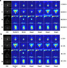
          The classification of extramural vascular invasion status using baseline magnetic resonance imaging in rectal cancer has gained significant attention as it is an important prognostic marker. Also, the accurate prediction of patients achieving complete response with primary staging MRI assists clinicians in determining subsequent treatment plans. Most studies utilised radiomics-based methods, requiring manually annotated segmentation and handcrafted features, which tend to generalise poorly. We retrospectively collected 509 patients from 9 centres, and proposed a fully automated pipeline for EMVI status classification and CR prediction with diffusion weighted imaging and T2-weighted imaging. We applied nnUNet, a self-configuring deep learning model, for tumour segmentation and employed learned multiple-level image features to train classification models, named MLNet. This ensures a more comprehensive representation of the tumour features, in terms of both fine-grained detail and global context. On external validation, MLNet, yielding similar AUCs as internal validation, outperformed 3D ResNet10, a deep neural network with ten layers designed for analysing spatiotemporal data, in both CR and EMVI tasks. For CR prediction, MLNet showed better results than the current state-of-the-art model using imaging and clinical features in the same external cohort. Our study demonstrated that incorporating multi-level image representations learned by a deep learning based tumour segmentation model on primary MRI improves the results of EMVI classification and CR prediction with good generalisation to external data. We observed variations in the contributions of individual feature maps to different classification tasks. This pipeline has the potential to be applied in clinical settings, particularly for EMVI classification.

          Related collections

          Most cited references34

          • Record: found
          • Abstract: found
          • Article: not found

          nnU-Net: a self-configuring method for deep learning-based biomedical image segmentation

          Biomedical imaging is a driver of scientific discovery and a core component of medical care and is being stimulated by the field of deep learning. While semantic segmentation algorithms enable image analysis and quantification in many applications, the design of respective specialized solutions is non-trivial and highly dependent on dataset properties and hardware conditions. We developed nnU-Net, a deep learning-based segmentation method that automatically configures itself, including preprocessing, network architecture, training and post-processing for any new task. The key design choices in this process are modeled as a set of fixed parameters, interdependent rules and empirical decisions. Without manual intervention, nnU-Net surpasses most existing approaches, including highly specialized solutions on 23 public datasets used in international biomedical segmentation competitions. We make nnU-Net publicly available as an out-of-the-box tool, rendering state-of-the-art segmentation accessible to a broad audience by requiring neither expert knowledge nor computing resources beyond standard network training.
            Bookmark
            • Record: found
            • Abstract: found
            • Article: not found

            Long-term outcome in patients with a pathological complete response after chemoradiation for rectal cancer: a pooled analysis of individual patient data.

            Locally advanced rectal cancer is usually treated with preoperative chemoradiation. After chemoradiation and surgery, 15-27% of the patients have no residual viable tumour at pathological examination, a pathological complete response (pCR). This study established whether patients with pCR have better long-term outcome than do those without pCR. In PubMed, Medline, and Embase we identified 27 articles, based on 17 different datasets, for long-term outcome of patients with and without pCR. 14 investigators agreed to provide individual patient data. All patients underwent chemoradiation and total mesorectal excision. Primary outcome was 5-year disease-free survival. Kaplan-Meier survival functions were computed and hazard ratios (HRs) calculated, with the Cox proportional hazards model. Subgroup analyses were done to test for effect modification by other predicting factors. Interstudy heterogeneity was assessed for disease-free survival and overall survival with forest plots and the Q test. 484 of 3105 included patients had a pCR. Median follow-up for all patients was 48 months (range 0-277). 5-year crude disease-free survival was 83.3% (95% CI 78.8-87.0) for patients with pCR (61/419 patients had disease recurrence) and 65.6% (63.6-68.0) for those without pCR (747/2263; HR 0.44, 95% CI 0.34-0.57; p<0.0001). The Q test and forest plots did not suggest significant interstudy variation. The adjusted HR for pCR for failure was 0.54 (95% CI 0.40-0.73), indicating that patients with pCR had a significantly increased probability of disease-free survival. The adjusted HR for disease-free survival for administration of adjuvant chemotherapy was 0.91 (95% CI 0.73-1.12). The effect of pCR on disease-free survival was not modified by other prognostic factors. Patients with pCR after chemoradiation have better long-term outcome than do those without pCR. pCR might be indicative of a prognostically favourable biological tumour profile with less propensity for local or distant recurrence and improved survival. None. Copyright 2010 Elsevier Ltd. All rights reserved.
              Bookmark
              • Record: found
              • Abstract: found
              • Article: found
              Is Open Access

              Deep Learning Techniques for Medical Image Segmentation: Achievements and Challenges

              Deep learning-based image segmentation is by now firmly established as a robust tool in image segmentation. It has been widely used to separate homogeneous areas as the first and critical component of diagnosis and treatment pipeline. In this article, we present a critical appraisal of popular methods that have employed deep-learning techniques for medical image segmentation. Moreover, we summarize the most common challenges incurred and suggest possible solutions.
                Bookmark

                Author and article information

                Contributors
                s.benson@nki.nl
                Journal
                NPJ Precis Oncol
                NPJ Precis Oncol
                NPJ Precision Oncology
                Nature Publishing Group UK (London )
                2397-768X
                22 January 2024
                22 January 2024
                2024
                : 8
                : 17
                Affiliations
                [1 ]Department of Radiology, The Netherlands Cancer Institute, ( https://ror.org/03xqtf034) Plesmanlaan 121, 1066 CX Amsterdam, The Netherlands
                [2 ]GROW School for Oncology and Developmental Biology, Maastricht University Medical Centre, P. Debyelaan 25, ( https://ror.org/02d9ce178) 66202 AZ Maastricht, The Netherlands
                [3 ]Department of Surgery, The Netherlands Cancer Institute, Plesmanlaan 121, ( https://ror.org/03xqtf034) 1066 CX Amsterdam, The Netherlands
                Author information
                http://orcid.org/0000-0002-3282-291X
                http://orcid.org/0000-0002-1671-9912
                Article
                516
                10.1038/s41698-024-00516-x
                10803303
                38253770
                14fd8196-4161-4583-8ed7-b70a7a1920e9
                © The Author(s) 2024

                Open Access This article is licensed under a Creative Commons Attribution 4.0 International License, which permits use, sharing, adaptation, distribution and reproduction in any medium or format, as long as you give appropriate credit to the original author(s) and the source, provide a link to the Creative Commons license, and indicate if changes were made. The images or other third party material in this article are included in the article’s Creative Commons license, unless indicated otherwise in a credit line to the material. If material is not included in the article’s Creative Commons license and your intended use is not permitted by statutory regulation or exceeds the permitted use, you will need to obtain permission directly from the copyright holder. To view a copy of this license, visit http://creativecommons.org/licenses/by/4.0/.

                History
                : 16 August 2023
                : 14 December 2023
                Funding
                Funded by: European Union’s Horizon 2020 research and innovation programme under the Marie Skłodowska-Curie grant agreement No 857894
                Categories
                Article
                Custom metadata
                © Springer Nature Limited 2024

                cancer imaging,risk factors
                cancer imaging, risk factors

                Comments

                Comment on this article