9
views
0
recommends
+1 Recommend
0 collections
    0
    shares
      • Record: found
      • Abstract: found
      • Article: found
      Is Open Access

      Dissecting cell-type-specific metabolism in pancreatic ductal adenocarcinoma

      research-article

      Read this article at

      Bookmark
          There is no author summary for this article yet. Authors can add summaries to their articles on ScienceOpen to make them more accessible to a non-specialist audience.

          Abstract

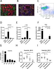
          Tumors are composed of many different cell types including cancer cells, fibroblasts, and immune cells. Dissecting functional metabolic differences between cell types within a mixed population can be challenging due to the rapid turnover of metabolites relative to the time needed to isolate cells. To overcome this challenge, we traced isotope-labeled nutrients into macromolecules that turn over more slowly than metabolites. This approach was used to assess differences between cancer cell and fibroblast metabolism in murine pancreatic cancer organoid-fibroblast co-cultures and tumors. Pancreatic cancer cells exhibited increased pyruvate carboxylation relative to fibroblasts, and this flux depended on both pyruvate carboxylase and malic enzyme 1 activity. Consequently, expression of both enzymes in cancer cells was necessary for organoid and tumor growth, demonstrating that dissecting the metabolism of specific cell populations within heterogeneous systems can identify dependencies that may not be evident from studying isolated cells in culture or bulk tissue.

          eLife digest

          Tumors contain a mixture of many different types of cells, including cancer cells and non-cancer cells. The interactions between these two groups of cells affect how the cancer cells use nutrients, which, in turn, affects how fast these cells grow and divide. Furthermore, different cell types may use nutrients in diverse ways to make other molecules – known as metabolites – that the cell needs to survive.

          Fibroblasts are a subset of non-cancer cells that are typically found in tumors and can help them form. Separating fibroblasts from cancer cells in a tumor takes a lot longer than the chemical reactions in each cell of the tumor that produce and use up nutrients, also known as the cell’s metabolism. Therefore, measuring the levels of glucose (the sugar that is the main energy source for cells) and other metabolites in each tumor cell after separating them does not necessarily provide accurate information about the tumor cell’s metabolism. This makes it difficult to study how cancer cells and fibroblasts use nutrients differently.

          Lau et al. have developed a strategy to study the metabolism of cancer cells and fibroblasts in tumors. Mice with tumors in their pancreas were provided glucose that had been labelled using biochemical techniques. As expected, when the cell processed the glucose, the label was transferred into metabolites that got used up very quickly. But the label also became incorporated into larger, more stable molecules, such as proteins. Unlike the small metabolites, these larger molecules do not change in the time it takes to separate the cancer cells from the fibroblasts.

          Lau et al. sorted cells from whole pancreatic tumors and analyzed large, stable molecules that can incorporate the label from glucose in cancer cells and fibroblasts. The experiments showed that, in cancer cells, these molecules were more likely to have labeling patterns that are characteristic of two specific enzymes called pyruvate carboxylase and malic enzyme 1. This suggests that these enzymes are more active in cancer cells. Lau et al. also found that pancreatic cancer cells needed these two enzymes to metabolize glucose and to grow into large tumors.

          Pancreatic cancer is one of the most lethal cancers and current therapies offer limited benefit to many patients. Therefore, it is important to develop new drugs to treat this disease. Understanding how cancer cells and non-cancer cells in pancreatic tumors use nutrients differently is important for developing drugs that only target cancer cells.

          Related collections

          Most cited references51

          • Record: found
          • Abstract: found
          • Article: not found

          Brain energy metabolism: focus on astrocyte-neuron metabolic cooperation.

          The energy requirements of the brain are very high, and tight regulatory mechanisms operate to ensure adequate spatial and temporal delivery of energy substrates in register with neuronal activity. Astrocytes-a type of glial cell-have emerged as active players in brain energy delivery, production, utilization, and storage. Our understanding of neuroenergetics is rapidly evolving from a "neurocentric" view to a more integrated picture involving an intense cooperativity between astrocytes and neurons. This review focuses on the cellular aspects of brain energy metabolism, with a particular emphasis on the metabolic interactions between neurons and astrocytes. Copyright © 2011 Elsevier Inc. All rights reserved.
            Bookmark
            • Record: found
            • Abstract: found
            • Article: found

            Oncogenic Kras Maintains Pancreatic Tumors through Regulation of Anabolic Glucose Metabolism

            Tumor maintenance relies on continued activity of driver oncogenes, although their rate-limiting role is highly context dependent. Oncogenic Kras mutation is the signature event in pancreatic ductal adenocarcinoma (PDAC), serving a critical role in tumor initiation. Here, an inducible Kras(G12D)-driven PDAC mouse model establishes that advanced PDAC remains strictly dependent on Kras(G12D) expression. Transcriptome and metabolomic analyses indicate that Kras(G12D) serves a vital role in controlling tumor metabolism through stimulation of glucose uptake and channeling of glucose intermediates into the hexosamine biosynthesis and pentose phosphate pathways (PPP). These studies also reveal that oncogenic Kras promotes ribose biogenesis. Unlike canonical models, we demonstrate that Kras(G12D) drives glycolysis intermediates into the nonoxidative PPP, thereby decoupling ribose biogenesis from NADP/NADPH-mediated redox control. Together, this work provides in vivo mechanistic insights into how oncogenic Kras promotes metabolic reprogramming in native tumors and illuminates potential metabolic targets that can be exploited for therapeutic benefit in PDAC. Copyright © 2012 Elsevier Inc. All rights reserved.
              Bookmark
              • Record: found
              • Abstract: found
              • Article: not found

              Lactate Metabolism in Human Lung Tumors

              Cancer cells consume glucose and secrete lactate in culture. It is unknown whether lactate contributes to energy metabolism in living tumors. We previously reported that human non-small cell lung cancers (NSCLC) oxidize glucose in the tricarboxylic acid (TCA) cycle. Here we show that lactate is also a TCA cycle carbon source for NSCLC. In human NSCLC, evidence of lactate utilization was most apparent in tumors with high 18 fluorodeoxyglucose uptake and aggressive oncological behavior. Infusing human NSCLC patients with 13 C-lactate revealed extensive labeling of TCA cycle metabolites. In mice, deleting monocarboxylate transporter-1 (MCT1) from tumor cells eliminated lactate-dependent metabolite labeling, confirming tumor-cell autonomous lactate uptake. Strikingly, directly comparing lactate and glucose metabolism in vivo indicated that lactate's contribution to the TCA cycle predominates. The data indicate that tumors, including bona fide human NSCLC, can use lactate as a fuel in vivo. Human non-small cell lung cancer preferentially utilizes lactate over glucose to fuel TCA cycle and sustain tumor metabolism in vivo.
                Bookmark

                Author and article information

                Contributors
                Role: Reviewing Editor
                Role: Senior Editor
                Journal
                eLife
                Elife
                eLife
                eLife
                eLife Sciences Publications, Ltd
                2050-084X
                10 July 2020
                2020
                : 9
                : e56782
                Affiliations
                [1 ]Koch Institute for Integrative Cancer Research and the Department of Biology at Massachusetts Institute of Technology CambridgeUnited States
                [2 ]Department of Biochemistry and Molecular Biology, University of Massachusetts, Amherst AmherstUnited States
                [3 ]Department of Biology and Biological Engineering, Chalmers University of Technology GothenburgSweden
                [4 ]Cold Spring Harbor Laboratory, Cold Spring Harbor New YorkUnited States
                [5 ]Lustgarten Foundation Pancreatic Cancer Research Laboratory, Cold Spring Harbor New YorkUnited States
                [6 ]Cancer Research United Kingdom Cambridge Institute, University of Cambridge CambridgeUnited Kingdom
                [7 ]Lewis-Sigler Institute for Integrative Genomics, Princeton University PrincetonUnited States
                [8 ]Department of Molecular Biology, Princeton University PrincetonUnited States
                [9 ]Department of Medicine, University of Cambridge CambridgeUnited Kingdom
                [10 ]Cambridge Institute for Therapeutic Immunology and Infectious Disease, University of Cambridge CambridgeUnited Kingdom
                [11 ]Department of Pathology, Massachusetts General Hospital BostonUnited States
                [12 ]Department of Medical Oncology, Dana-Farber Cancer Institute BostonUnited States
                UT Southwestern Medical Center United States
                Memorial Sloan Kettering Cancer Center United States
                UT Southwestern Medical Center United States
                UT Southwestern Medical Center United States
                Princeton University United States
                Author information
                https://orcid.org/0000-0003-4250-7355
                http://orcid.org/0000-0001-9881-6232
                http://orcid.org/0000-0002-8607-1787
                http://orcid.org/0000-0002-3318-1851
                https://orcid.org/0000-0002-6702-4192
                Article
                56782
                10.7554/eLife.56782
                7406355
                32648540
                145ed62a-2bf9-471a-be0b-017216bda53d
                © 2020, Lau et al

                This article is distributed under the terms of the Creative Commons Attribution License, which permits unrestricted use and redistribution provided that the original author and source are credited.

                History
                : 09 March 2020
                : 09 July 2020
                Funding
                Funded by: FundRef http://dx.doi.org/10.13039/100001021, Damon Runyon Cancer Research Foundation;
                Award ID: DRG-2241-15
                Award Recipient :
                Funded by: FundRef http://dx.doi.org/10.13039/100001021, Damon Runyon Cancer Research Foundation;
                Award ID: DRG-2367-19
                Award Recipient :
                Funded by: FundRef http://dx.doi.org/10.13039/100001021, Damon Runyon Cancer Research Foundation;
                Award ID: DRG-2299-17
                Award Recipient :
                Funded by: FundRef http://dx.doi.org/10.13039/100000054, National Cancer Institute;
                Award ID: K99CA234221
                Award Recipient :
                Funded by: FundRef http://dx.doi.org/10.13039/100000002, National Institutes of Health;
                Award ID: T32GM007287
                Award Recipient :
                Funded by: FundRef http://dx.doi.org/10.13039/100001033, Jane Coffin Childs Memorial Fund for Medical Research;
                Award Recipient :
                Funded by: FundRef http://dx.doi.org/10.13039/501100001729, Swedish Foundation for Strategic Research;
                Award Recipient :
                Funded by: FundRef http://dx.doi.org/10.13039/501100004063, Knut and Alice Wallenberg Foundation;
                Award Recipient :
                Funded by: FundRef http://dx.doi.org/10.13039/100008483, Barbro Osher Pro Suecia Foundation;
                Award Recipient :
                Funded by: FundRef http://dx.doi.org/10.13039/100000054, National Cancer Institute;
                Award ID: U54CA163109
                Award Recipient :
                Funded by: FundRef http://dx.doi.org/10.13039/100004412, Human Frontier Science Program;
                Award ID: LT000195/2015-L
                Award Recipient :
                Funded by: FundRef http://dx.doi.org/10.13039/501100003043, EMBO;
                Award ID: ALTF 1203-2014
                Award Recipient :
                Funded by: FundRef http://dx.doi.org/10.13039/100000011, Howard Hughes Medical Institute;
                Award Recipient :
                Funded by: FundRef http://dx.doi.org/10.13039/501100000265, MRC;
                Award ID: CSF MR/P008801/1
                Award Recipient :
                Funded by: NHSBT;
                Award ID: WPA15-02
                Award Recipient :
                Funded by: NIHR Cambridge BRC;
                Award Recipient :
                Funded by: FundRef http://dx.doi.org/10.13039/100000002, National Institutes of Health;
                Award ID: R01CA211184
                Award Recipient :
                Funded by: FundRef http://dx.doi.org/10.13039/100000002, National Institutes of Health;
                Award ID: R01CA034992
                Award Recipient :
                Funded by: FundRef http://dx.doi.org/10.13039/100005979, Lustgarten Foundation;
                Award Recipient :
                Funded by: FundRef http://dx.doi.org/10.13039/100009730, Stand Up To Cancer;
                Award Recipient :
                Funded by: MIT Center for Precision Cancer Medicine;
                Award Recipient :
                Funded by: Ludwig Center at MIT;
                Award Recipient :
                Funded by: Emerald Foundation;
                Award Recipient :
                Funded by: FundRef http://dx.doi.org/10.13039/100000054, National Cancer Institute;
                Award ID: R01CA168653
                Award Recipient :
                Funded by: FundRef http://dx.doi.org/10.13039/100000054, National Cancer Institute;
                Award ID: R01CA201276
                Award Recipient :
                Funded by: FundRef http://dx.doi.org/10.13039/100000054, National Cancer Institute;
                Award ID: R35CA242379
                Award Recipient :
                Funded by: FundRef http://dx.doi.org/10.13039/100000054, National Cancer Institute;
                Award ID: P30CA14051
                Award Recipient :
                The funders had no role in study design, data collection and interpretation, or the decision to submit the work for publication.
                Categories
                Research Article
                Cancer Biology
                Custom metadata
                Isotope tracing into macromolecules enables functional analysis of metabolism in specific cell populations in tumors and provides insight into metabolic differences between cancer and stromal cells in their endogenous microenvironment.

                Life sciences
                pancreatic cancer,organoid culture,malic enzyme 1,pdac,pyruvate carboxylase,metabolic heterogeneity,mouse

                Comments

                Comment on this article