8
views
0
recommends
+1 Recommend
0 collections
    0
    shares
      • Record: found
      • Abstract: found
      • Article: found
      Is Open Access

      OCTA in neurodegenerative optic neuropathies: emerging biomarkers at the eye–brain interface

      review-article

      Read this article at

      Bookmark
          There is no author summary for this article yet. Authors can add summaries to their articles on ScienceOpen to make them more accessible to a non-specialist audience.

          Abstract

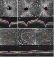
          OCTA imaging in optic neuropathies.

          Related collections

          Most cited references84

          • Record: found
          • Abstract: not found
          • Article: not found

          Alzheimer's disease and Parkinson's disease.

            Bookmark
            • Record: found
            • Abstract: found
            • Article: found
            Is Open Access

            A review of optical coherence tomography angiography (OCTA)

            Optical coherence tomography angiography (OCTA) is a new, non-invasive imaging technique that generates volumetric angiography images in a matter of seconds. This is a nascent technology with a potential wide applicability for retinal vascular disease. At present, level 1 evidence of the technology’s clinical applications doesn’t exist. In this paper, we introduce the technology, review the available English language publications regarding OCTA, and compare it with the current angiographic gold standards, fluorescein angiography (FA) and indocyanine green angiography (ICGA). Finally we summarize its potential application to retinal vascular diseases. OCTA is quick and non-invasive, and provides volumetric data with the clinical capability of specifically localizing and delineating pathology along with the ability to show both structural and blood flow information in tandem. Its current limitations include a relatively small field of view, inability to show leakage, and proclivity for image artifact due to patient movement/blinking. Published studies hint at OCTA’s potential efficacy in the evaluation of common ophthalmologic diseases such age related macular degeneration (AMD), diabetic retinopathy, artery and vein occlusions, and glaucoma. OCTA can detect changes in choroidal blood vessel flow and can elucidate the presence of choroidal neovascularization (CNV) in a variety of conditions but especially in AMD. It provides a highly detailed view of the retinal vasculature, which allows for accurate delineation of the foveal avascular zone (FAZ) in diabetic eyes and detection of subtle microvascular abnormalities in diabetic and vascular occlusive eyes. Optic disc perfusion in glaucomatous eyes is notable as well on OCTA. Further studies are needed to more definitively determine OCTA’s utility in the clinical setting and to establish if this technology may offer a non-invasive option of visualizing the retinal vasculature in detail.
              Bookmark
              • Record: found
              • Abstract: found
              • Article: not found

              Microvascular network alterations in the retina of patients with Alzheimer's disease.

              Although cerebral small-vessel disease has been implicated in the development of Alzheimer's disease (AD), the cerebral microcirculation is difficult to visualize directly in vivo. Because the retina provides a noninvasive window to assess the microcirculation, we determined whether quantitatively measured retinal microvascular parameters are associated with AD. We conducted a case-control study (case:control matching ≈ 1:2). Retinal photographs were analyzed using a computer program, and a spectrum of quantitative retinal microvascular parameters (caliber, fractal dimension, tortuosity, and bifurcation) were measured. Logistic regression models were used to compute the odds ratio (OR) and 95% confidence interval for AD adjusting for age, gender, ethnicity, smoking, hypertension, diabetes, hypercholesterolemia, and history of myocardial infarction. We included 136 demented patients with AD and 290 age-gender-race-matched controls. Persons with narrower venular caliber (OR per standard deviation [SD] decrease, 2.01 [1.27-3.19]), decreased arteriolar and venular fractal dimension (OR per SD decrease 1.35 [1.08-1.68], 1.47 [1.17-1.84], respectively) and increased arteriolar and venular tortuosity (OR per SD increase, 1.84 [1.40-2.31], 1.94 [1.48-2.53], respectively) were more likely to have AD. These associations still persisted when only AD cases without a history of cerebrovascular disease were included. Patients with AD have altered microvascular network in the retina (narrower retinal venules and a sparser and more tortuous retinal vessels) compared with matched nondemented controls. These changes in retinal microvasculature may reflect similar pathophysiological processes in cerebral microvasculature in the brains of patients with AD. Copyright © 2014 The Alzheimer's Association. Published by Elsevier Inc. All rights reserved.
                Bookmark

                Author and article information

                Contributors
                Journal
                Ther Adv Ophthalmol
                Ther Adv Ophthalmol
                OED
                spoed
                Therapeutic Advances in Ophthalmology
                SAGE Publications (Sage UK: London, England )
                2515-8414
                27 August 2020
                Jan-Dec 2020
                : 12
                : 2515841420950508
                Affiliations
                [1-2515841420950508]Department of Ophthalmology and Visual Sciences, University of Maryland Eye Associates, University of Maryland Medical Center and University of Maryland School of Medicine, 419 W. Redwood St., Baltimore, MD 21201, USA
                [2-2515841420950508]Department of Ophthalmology and Visual Sciences, University of Maryland School of Medicine, Baltimore, MD, USA
                [3-2515841420950508]Doheny Eye Center, Los Angeles, CA, USA; Department of Ophthalmology, David Geffen School of Medicine, University of California, Los Angeles, Los Angeles, CA, USA
                [4-2515841420950508]Department of Ophthalmology and Visual Sciences, University of Maryland School of Medicine, Baltimore, MD, USA
                Author notes
                Author information
                https://orcid.org/0000-0002-7556-0155
                Article
                10.1177_2515841420950508
                10.1177/2515841420950508
                7457690
                32923939
                1389a617-3399-4c8f-9e1d-11cbdfdc203e
                © The Author(s), 2020

                This article is distributed under the terms of the Creative Commons Attribution-NonCommercial 4.0 License ( https://creativecommons.org/licenses/by-nc/4.0/) which permits non-commercial use, reproduction and distribution of the work without further permission provided the original work is attributed as specified on the SAGE and Open Access page ( https://us.sagepub.com/en-us/nam/open-access-at-sage).

                History
                : 13 March 2020
                : 13 July 2020
                Categories
                Recent Advances in Ocular Imaging
                Review
                Custom metadata
                January-December 2020
                ts1

                alzheimer’s disease,dominant optic atrophy,erythrocyte mediated angiography,glaucoma,leber’s hereditary optic neuropathy,optic neuropathy,optical coherence tomography angiography,parkinson’s disease,retinal blood flow,schizophrenia,vasomotion,wolfram syndrome

                Comments

                Comment on this article