19
views
0
recommends
+1 Recommend
0 collections
    0
    shares
      • Record: found
      • Abstract: found
      • Article: found
      Is Open Access

      KRAS‐G12D mutation drives immune suppression and the primary resistance of anti‐PD‐1/PD‐L1 immunotherapy in non‐small cell lung cancer

      research-article

      Read this article at

      Bookmark
          There is no author summary for this article yet. Authors can add summaries to their articles on ScienceOpen to make them more accessible to a non-specialist audience.

          Abstract

          Background

          Although immune checkpoint inhibitors (ICIs) against programmed cell death protein 1 (PD‐1) and its ligand PD‐L1 have demonstrated potency towards treating patients with non‐small cell lung carcinoma (NSCLC), the potential association between Kirsten rat sarcoma viral oncogene homolog ( KRAS) oncogene substitutions and the efficacy of ICIs remains unclear. In this study, we aimed to find point mutations in the KRAS gene resistant to ICIs and elucidate resistance mechanism.

          Methods

          The association between KRAS variant status and the efficacy of ICIs was explored with a clinical cohort ( n = 74), and confirmed with a mouse model. In addition, the tumor immune microenvironment (TIME) of KRAS‐mutant NSCLC, such as CD8 + tumor‐infiltrating lymphocytes (TILs) and PD‐L1 level, was investigated. Cell lines expressing classic KRAS substitutions were used to explore signaling pathway activation involved in the formation of TIME. Furthermore, interventions that improved TIME were developed to increase responsiveness to ICIs.

          Results

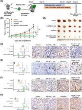
          We observed the inferior efficacy of ICIs in KRAS‐G12D‐mutant NSCLC. Based upon transcriptome data and immunostaining results from KRAS‐mutant NSCLC, KRAS‐G12D point mutation negatively correlated with PD‐L1 level and secretion of chemokines CXCL10/CXCL11 that led to a decrease in CD8 + TILs, which in turn yielded an immunosuppressive TIME. The analysis of cell lines overexpressing classic KRAS substitutions further revealed that KRAS‐G12D mutation suppressed PD‐L1 level via the P70S6K/PI3K/AKT axis and reduced CXCL10/CXCL11 levels by down‐regulating high mobility group protein A2 (HMGA2) level. Notably, paclitaxel, a chemotherapeutic agent, upregulated HMGA2 level, and in turn, stimulated the secretion of CXCL10/CXCL11. Moreover, PD‐L1 blockade combined with paclitaxel significantly suppressed tumor growth compared with PD‐L1 inhibitor monotherapy in a mouse model with KRAS‐G12D‐mutant lung adenocarcinoma. Further analyses revealed that the combined treatment significantly enhanced the recruitment of CD8 + TILs via the up‐regulation of CXCL10/CXCL11 levels. Results of clinical study also revealed the superior efficacy of chemo‐immunotherapy in patients with KRAS‐G12D‐mutant NSCLC compared with ICI monotherapy.

          Conclusions

          Our study elucidated the molecular mechanism by which KRAS‐G12D mutation drives immunosuppression and enhances resistance of ICIs in NSCLC. Importantly, our findings demonstrate that ICIs in combination with chemotherapy may be more effective in patients with KRAS‐G12D‐mutant NSCLC.

          Related collections

          Most cited references67

          • Record: found
          • Abstract: found
          • Article: found
          Is Open Access

          Moderated estimation of fold change and dispersion for RNA-seq data with DESeq2

          In comparative high-throughput sequencing assays, a fundamental task is the analysis of count data, such as read counts per gene in RNA-seq, for evidence of systematic changes across experimental conditions. Small replicate numbers, discreteness, large dynamic range and the presence of outliers require a suitable statistical approach. We present DESeq2, a method for differential analysis of count data, using shrinkage estimation for dispersions and fold changes to improve stability and interpretability of estimates. This enables a more quantitative analysis focused on the strength rather than the mere presence of differential expression. The DESeq2 package is available at http://www.bioconductor.org/packages/release/bioc/html/DESeq2.html. Electronic supplementary material The online version of this article (doi:10.1186/s13059-014-0550-8) contains supplementary material, which is available to authorized users.
            Bookmark
            • Record: found
            • Abstract: found
            • Article: not found

            Cancer statistics, 2019

            Each year, the American Cancer Society estimates the numbers of new cancer cases and deaths that will occur in the United States and compiles the most recent data on cancer incidence, mortality, and survival. Incidence data, available through 2015, were collected by the Surveillance, Epidemiology, and End Results Program; the National Program of Cancer Registries; and the North American Association of Central Cancer Registries. Mortality data, available through 2016, were collected by the National Center for Health Statistics. In 2019, 1,762,450 new cancer cases and 606,880 cancer deaths are projected to occur in the United States. Over the past decade of data, the cancer incidence rate (2006-2015) was stable in women and declined by approximately 2% per year in men, whereas the cancer death rate (2007-2016) declined annually by 1.4% and 1.8%, respectively. The overall cancer death rate dropped continuously from 1991 to 2016 by a total of 27%, translating into approximately 2,629,200 fewer cancer deaths than would have been expected if death rates had remained at their peak. Although the racial gap in cancer mortality is slowly narrowing, socioeconomic inequalities are widening, with the most notable gaps for the most preventable cancers. For example, compared with the most affluent counties, mortality rates in the poorest counties were 2-fold higher for cervical cancer and 40% higher for male lung and liver cancers during 2012-2016. Some states are home to both the wealthiest and the poorest counties, suggesting the opportunity for more equitable dissemination of effective cancer prevention, early detection, and treatment strategies. A broader application of existing cancer control knowledge with an emphasis on disadvantaged groups would undoubtedly accelerate progress against cancer.
              Bookmark
              • Record: found
              • Abstract: found
              • Article: not found

              Cancer immunotherapy using checkpoint blockade

              The release of negative regulators of immune activation (immune checkpoints) that limit antitumor responses has resulted in unprecedented rates of long-lasting tumor responses in patients with a variety of cancers. This can be achieved by antibodies blocking the cytotoxic T lymphocyte antigen-4 (CTLA-4) or the programmed death-1 (PD-1) pathway, either alone or in combination. The main premise for inducing an immune response is the pre-existence of antitumor T cells that were limited by specific immune checkpoints. Most patients who have tumor responses maintain long lasting disease control, yet one third of patients relapse. Mechanisms of acquired resistance are currently poorly understood, but evidence points to alterations that converge on the antigen presentation and interferon gamma signaling pathways. New generation combinatorial therapies may overcome resistance mechanisms to immune checkpoint therapy.
                Bookmark

                Author and article information

                Contributors
                sunnan@vip.126.com
                prof.jiehe@gmail.com
                Journal
                Cancer Commun (Lond)
                Cancer Commun (Lond)
                10.1002/(ISSN)2523-3548
                CAC2
                Cancer Communications
                John Wiley and Sons Inc. (Hoboken )
                2523-3548
                11 July 2022
                September 2022
                : 42
                : 9 ( doiID: 10.1002/cac2.v42.9 )
                : 828-847
                Affiliations
                [ 1 ] Department of Thoracic Surgery National Cancer Center/National Clinical Research Center for Cancer/Cancer Hospital Chinese Academy of Medical Sciences and Peking Union Medical College Beijing 100021 P. R. China
                [ 2 ] State Key Laboratory of Molecular Oncology National Cancer Center/National Clinical Research Center for Cancer/Cancer Hospital Chinese Academy of Medical Sciences and Peking Union Medical College Beijing 100021 P. R. China
                [ 3 ] Department of Medical Oncology National Cancer Center/National Clinical Research Center for Cancer/Cancer Hospital Chinese Academy of Medical Sciences and Peking Union Medical College Beijing 100021 P. R. China
                [ 4 ] Department of Comprehensive Oncology National Cancer Center/National Clinical Research Center for Cancer/Cancer Hospital Chinese Academy of Medical Sciences and Peking Union Medical College Beijing 100021 P. R. China
                [ 5 ] Department of Pathology National Cancer Center/National Clinical Research Center for Cancer/Cancer Hospital Chinese Academy of Medical Sciences and Peking Union Medical College Beijing 100021 P. R. China
                Author notes
                [*] [* ] Correspondence

                Jie He, Department of Thoracic Surgery, National Cancer Center/National Clinical Research Center for Cancer/Cancer Hospital, Chinese Academy of Medical Sciences and Peking Union Medical College, Beijing 100021, P. R. China.

                Email: prof.jiehe@ 123456gmail.com

                Nan Sun, State Key Laboratory of Molecular Oncology, National Cancer Center/National Clinical Research Center for Cancer/Cancer Hospital, Chinese Academy of Medical Sciences and Peking Union Medical College, Beijing 100021, P. R. China.

                Email: sunnan@ 123456vip.126.com

                Author information
                https://orcid.org/0000-0002-5086-4654
                https://orcid.org/0000-0002-1743-6383
                https://orcid.org/0000-0002-0285-5403
                Article
                CAC212327
                10.1002/cac2.12327
                9456691
                35811500
                137a29a5-7c31-4228-a159-6a3c95dc77f0
                © 2022 The Authors. Cancer Communications published by John Wiley & Sons Australia, Ltd. on behalf of Sun Yat‐sen University Cancer Center.

                This is an open access article under the terms of the http://creativecommons.org/licenses/by-nc-nd/4.0/ License, which permits use and distribution in any medium, provided the original work is properly cited, the use is non‐commercial and no modifications or adaptations are made.

                History
                : 19 April 2022
                : 26 January 2022
                : 14 June 2022
                Page count
                Figures: 8, Tables: 0, Pages: 20, Words: 10433
                Funding
                Funded by: National Natural Science Foundation of China , doi 10.13039/501100001809;
                Award ID: 82072590
                Award ID: 81502514
                Award ID: 81802299
                Funded by: Graduate Innovation Funds of Peking Union Medical College
                Award ID: 2019‐1002‐06
                Funded by: National Key Basic Research Development Plan
                Award ID: 2018YFC1312105
                Funded by: National Key R&D Program of China
                Award ID: 2018YFC1312100
                Award ID: 2018YFC1312102
                Funded by: Fundamental Research Funds for the Central Universities , doi 10.13039/501100012226;
                Award ID: 3332018070
                Funded by: CAMS Innovation Fund for Medical Sciences
                Award ID: 2016‐I2M‐1‐001
                Award ID: 2017‐I2M‐1‐005
                Categories
                Original Article
                Original Articles
                Custom metadata
                2.0
                September 2022
                Converter:WILEY_ML3GV2_TO_JATSPMC version:6.1.8 mode:remove_FC converted:08.09.2022

                kras‐g12d,non‐small cell lung carcinoma,immunotherapy,pd‐l1,tumor‐infiltrating lymphocyte,chemo‐immunotherapy

                Comments

                Comment on this article