3
views
0
recommends
+1 Recommend
0 collections
    0
    shares
      • Record: found
      • Abstract: found
      • Article: found
      Is Open Access

      Femtosecond pulsed laser photodynamic therapy activates melanin and eradicates malignant melanoma

      research-article

      Read this article at

      Bookmark
          There is no author summary for this article yet. Authors can add summaries to their articles on ScienceOpen to make them more accessible to a non-specialist audience.

          Significance

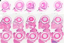
          While effective in non-pigmented tumors, photodynamic therapy (PDT) has failed in treating melanoma due to the high light attenuation by melanin. Here, an effective approach for ocular melanoma is proposed using the concept of melanin-mediated multi-photon PDT. Experiments were performed comparing cytotoxicity in pigmented versus nonpigmented melanoma cells treated by 1- versus 2-photon PDT using low versus high 2-photon cross-section photosensitizers. The unexpected results show that, under femtosecond pulsed laser irradiation, melanin can absorb 2 and 3 photons and transfer the energy to the photosensitizer. The resulting therapeutic efficacy is demonstrated in vivo in a murine model of conjunctival melanoma, achieving complete tumor eradication and showing the potential of this approach as a minimally invasive therapeutic option.

          Abstract

          Photodynamic therapy (PDT) relies on a series of photophysical and photochemical reactions leading to cell death. While effective for various cancers, PDT has been less successful in treating pigmented melanoma due to high light absorption by melanin. Here, this limitation is addressed by 2-photon excitation of the photosensitizer (2p-PDT) using ~100 fs pulses of near-infrared laser light. A critical role of melanin in enabling rather than hindering 2p-PDT is elucidated using pigmented and non-pigmented murine melanoma clonal cell lines in vitro. The photocytotoxicities were compared between a clinical photosensitizer (Visudyne) and a porphyrin dimer (Oxdime) with ~600-fold higher σ 2p value. Unexpectedly, while the 1p-PDT responses are similar in both cell lines, 2p activation is much more effective in killing pigmented than non-pigmented cells, suggesting a dominant role of melanin 2p-PDT. The potential for clinical translational is demonstrated in a conjunctival melanoma model in vivo, where complete eradication of small tumors was achieved. This work elucidates the melanin contribution in multi-photon PDT enabling significant advancement of light-based treatments that have previously been considered unsuitable in pigmented tumors.

          Related collections

          Most cited references46

          • Record: found
          • Abstract: found
          • Article: not found

          Uveal melanoma: trends in incidence, treatment, and survival.

          To determine trends in incidence, treatment, and survival with primary uveal melanoma in the United States over a 36-year period from 1973 to 2008. Systematic review of existing databases. A total of 4070 patients with primary uveal melanoma (International Classification of Disease for Oncology [ICD-O-2] codes C69.3 [choroid], C69.4 [ciliary body and iris], and C69.2 [retina]) derived from the Surveillance, Epidemiology, and End Results (SEER) program database in the United States from 1973 to 2008. The significance of trends in age-adjusted incidence, treatment, and 5-year relative survival rates were determined using chi-square testing and 95% confidence intervals (CIs). Age-adjusted incidence, form of treatment (surgery, radiation, or both), and 5-year relative survival rates. There were 4070 cases of uveal melanoma representing 3.1% of all recorded cases of melanoma. The majority of cases (98.3%) were reported by hospital inpatient/outpatient clinics. Histopathologic confirmation was available in 2804 cases (72.1% for all years). The mean age-adjusted incidence of uveal melanoma in the United States was 5.1 per million (95% CI, 4.8-5.3). The majority of cases (97.8%) occurred in the white population. There was a statistically significant variation of age-adjusted incidence between sexes (male = 5.8, 95% CI, 5.5-6.2; and female = 4.4, 95% CI, 4.2-4.7). A decreasing trend was observed in patients treated with surgery alone (93.8% for 1973-1975 vs. 28.3% for 2006-2008), whereas a corresponding increase was seen in those treated with radiation (1.8% for 1973-1975 vs. 62.5% for 2006-2008). No change in the 5-year relative survival rate (81.6%) was observed from 1973 to 2008. The age-adjusted incidence of uveal melanoma (5.1 per million) has remained unchanged from 1973 to 2008. Despite a shift toward more conservative treatments, survival has not improved during this time period. Copyright © 2011 American Academy of Ophthalmology. Published by Elsevier Inc. All rights reserved.
            Bookmark
            • Record: found
            • Abstract: found
            • Article: found
            Is Open Access

            Treatment of uveal melanoma: where are we now?

            Uveal melanoma, a rare subset of melanoma, is the most common primary intraocular malignancy in adults. Despite effective primary therapy, nearly 50% of patients will develop metastatic disease. Outcomes for those with metastatic disease remain dismal due to a lack of effective therapies. The unique biology and immunology of uveal melanoma necessitates the development of dedicated management and treatment approaches. Ongoing efforts seek to optimize the efficacy of targeted therapy and immunotherapy in both the adjuvant and metastatic setting. This review provides a comprehensive, updated overview of disease biology and risk stratification, the management of primary disease, options for adjuvant therapy, and the current status of treatment strategies for metastatic disease.
              Bookmark
              • Record: found
              • Abstract: found
              • Article: not found

              Two-photon excitation nanoparticles for photodynamic therapy.

              Two-photon excitation (TPE) nanoparticle-based photosensitizers (PSs) that combine the advantages of TPE and nanotechnology have emerged as attractive therapeutic agents for near-infrared red (NIR) light excited photodynamic therapy (PDT) for cancer treatment. TPE PDT is characterized by nonlinear absorption of two relatively low-energy photons of NIR light with the resulting emission of high-energy visible light. This high-energy light can sensitize oxygen to produce cytotoxic reactive oxygen species (ROS) and singlet oxygen ((1)O2) which can kill cancer cells. The long-wavelength light used to excite TPE NPs allows for deeper tissue penetration to achieve efficient PDT of deep-seated tumors. Moreover, TPE nanoparticles normally have large two-photon absorption (TPA) cross-sections, which hold great potential as efficient two-photon donors in PDT. In this review, we will summarize the recent advances made in the development of TPE nanoparticles for cancer PDT. Five different TPE nanoparticles, including quantum dots (QDs), carbon nanomaterials, silica nanoparticles, gold nanomaterials, and polymer nanoparticles, are summarized in detail, and the existing challenges as well as the future perspectives are also discussed.
                Bookmark

                Author and article information

                Contributors
                Journal
                Proc Natl Acad Sci U S A
                Proc Natl Acad Sci U S A
                PNAS
                Proceedings of the National Academy of Sciences of the United States of America
                National Academy of Sciences
                0027-8424
                1091-6490
                29 March 2024
                2 April 2024
                29 March 2024
                : 121
                : 14
                : e2316303121
                Affiliations
                [1] aDepartment of Cancer Biology and Imaging, Princess Margaret Cancer Center, University Health Network , Toronto, ON M5G 1L7, Canada
                [2] bDepartamento de Fisica e Ciencia dos Materiais, São Carlos Institute of Physics, University of São Paulo , Sao Carlos 13566-590, Brazil
                [3] cDepartments of Ophthalmology & Vision Sciences, St. Michael’s Hospital, University of Toronto , Toronto, ON M5B 1W8, Canada
                [4] dFaculty of Medicine, Department of Ophthalmology, Ophthalmology and Visual Sciences, University of British Columbia , Vancouver, BC V5Z 3N9, Canada
                [5] eDepartment of Biomedical Engineering, Texas A&M University , College Station, TX 77843
                [6] fFaculty of Medicine, Department of Medical Biophysics, University of Toronto , Toronto, ON M5G 1L7, Canada
                Author notes
                1To whom correspondence may be addressed. Email: vander@ 123456ifsc.usp.ca , cristina@ 123456ifsc.usp.br , or brian.wilson@ 123456uhn.ca .

                Contributed by Vanderlei S. Bagnato; received September 19, 2023; accepted January 14, 2024; reviewed by Keith Cengel and Angelika Rueck

                Author information
                https://orcid.org/0000-0003-4833-239X
                https://orcid.org/0000-0001-7175-5337
                Article
                202316303
                10.1073/pnas.2316303121
                10998568
                38551838
                12a198ee-6378-4b47-9cde-b0646a5aadb2
                Copyright © 2024 the Author(s). Published by PNAS.

                This open access article is distributed under Creative Commons Attribution-NonCommercial-NoDerivatives License 4.0 (CC BY-NC-ND).

                History
                : 19 September 2023
                : 14 January 2024
                Page count
                Pages: 9, Words: 4569
                Funding
                Funded by: Fundação de Amparo à Pesquisa do Estado de São Paulo (FAPESP), FundRef 501100001807;
                Award ID: 2013-07276-1
                Award Recipient : Vanderlei S. Bagnato
                Funded by: Conselho Nacional de Desenvolvimento Científico e Tecnológico (CNPq), FundRef 501100003593;
                Award ID: 305795/2016-3
                Award Recipient : Cristina Kurachi
                Funded by: CNPq | Ciência sem Fronteiras (CsF), FundRef 501100017564;
                Award ID: N/A
                Award Recipient : Layla Pires
                Funded by: Coordenação de Aperfeiçoamento de Pessoal de Nível Superior (CAPES), FundRef 501100002322;
                Award ID: N/A
                Award Recipient : Layla Pires
                Funded by: Universidade de Sao Paulo - University of Toronto Joint Program;
                Award ID: N/A
                Award Recipient : Layla Pires Award Recipient : Shireen Khattak Award Recipient : Yeni Yucel Award Recipient : Vanderlei S. Bagnato Award Recipient : Brian C Wilson
                Funded by: Banting Fellowship from the Canadian Institutes of Health Research;
                Award ID: N/A
                Award Recipient : Layla Pires Award Recipient : Shireen Khattak Award Recipient : Yeni Yucel Award Recipient : Vanderlei S. Bagnato Award Recipient : Brian C Wilson
                Funded by: Princess Margaret Cancer Centre Foundation Invest-in-Research Fund;
                Award ID: N/A
                Award Recipient : Layla Pires Award Recipient : Shireen Khattak Award Recipient : Yeni Yucel Award Recipient : Vanderlei S. Bagnato Award Recipient : Brian C Wilson
                Funded by: Princess Margaret Cancer Centre Foundation Excellence Fellowship Award;
                Award ID: N/A
                Award Recipient : Layla Pires Award Recipient : Shireen Khattak Award Recipient : Yeni Yucel Award Recipient : Vanderlei S. Bagnato Award Recipient : Brian C Wilson
                Funded by: Henry Farrugia Research Fund;
                Award ID: N/A
                Award Recipient : Layla Pires Award Recipient : Shireen Khattak Award Recipient : Yeni Yucel Award Recipient : Vanderlei S. Bagnato Award Recipient : Brian C Wilson
                Funded by: Vision Science Research Program Award;
                Award ID: N/A
                Award Recipient : Layla Pires Award Recipient : Shireen Khattak Award Recipient : Yeni Yucel Award Recipient : Vanderlei S. Bagnato Award Recipient : Brian C Wilson
                Funded by: Cancer Prevention and Research Institute of Texas (CPRIT), FundRef 100004917;
                Award ID: M20301556
                Award Recipient : Vanderlei S. Bagnato
                Funded by: Governor's University Research Initiative;
                Award ID: M230930
                Award Recipient : Layla Pires Award Recipient : Shireen Khattak Award Recipient : Yeni Yucel Award Recipient : Vanderlei S. Bagnato Award Recipient : Brian C Wilson
                Categories
                research-article, Research Article
                biophys-bio, Biophysics and Computational Biology
                408
                Biological Sciences
                Biophysics and Computational Biology

                photodynamic therapy,multiphoton,ocular melanoma,melanoma,melanin

                Comments

                Comment on this article