5
views
0
recommends
+1 Recommend
0 collections
    0
    shares
      • Record: found
      • Abstract: found
      • Article: found
      Is Open Access

      A Comparison of Static and Dynamic Functional Connectivities for Identifying Subjects and Biological Sex Using Intrinsic Individual Brain Connectivity

      research-article
      ,
      Scientific Reports
      Nature Publishing Group UK

      Read this article at

      Bookmark
          There is no author summary for this article yet. Authors can add summaries to their articles on ScienceOpen to make them more accessible to a non-specialist audience.

          Abstract

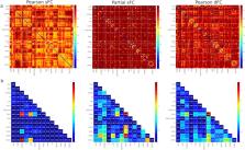
          Functional magnetic resonance imaging has revealed correlated activities in brain regions even in the absence of a task. Initial studies assumed this resting-state functional connectivity (FC) to be stationary in nature, but recent studies have modeled these activities as a dynamic network. Dynamic spatiotemporal models better model the brain activities, but are computationally more involved. A comparison of static and dynamic FCs was made to quantitatively study their efficacies in identifying intrinsic individual connectivity patterns using data from the Human Connectome Project. Results show that the intrinsic individual brain connectivity pattern can be used as a ‘fingerprint’ to distinguish among and identify subjects and is more accurately captured with partial correlation and assuming static FC. It was also seen that the intrinsic individual brain connectivity patterns were invariant over a few months. Additionally, biological sex identification was successfully performed using the intrinsic individual connectivity patterns, and group averages of male and female FC matrices. Edge consistency, edge variability and differential power measures were used to identify the major resting-state networks involved in identifying subjects and their sex.

          Related collections

          Most cited references25

          • Record: found
          • Abstract: found
          • Article: not found

          Network modelling methods for FMRI.

          There is great interest in estimating brain "networks" from FMRI data. This is often attempted by identifying a set of functional "nodes" (e.g., spatial ROIs or ICA maps) and then conducting a connectivity analysis between the nodes, based on the FMRI timeseries associated with the nodes. Analysis methods range from very simple measures that consider just two nodes at a time (e.g., correlation between two nodes' timeseries) to sophisticated approaches that consider all nodes simultaneously and estimate one global network model (e.g., Bayes net models). Many different methods are being used in the literature, but almost none has been carefully validated or compared for use on FMRI timeseries data. In this work we generate rich, realistic simulated FMRI data for a wide range of underlying networks, experimental protocols and problematic confounds in the data, in order to compare different connectivity estimation approaches. Our results show that in general correlation-based approaches can be quite successful, methods based on higher-order statistics are less sensitive, and lag-based approaches perform very poorly. More specifically: there are several methods that can give high sensitivity to network connection detection on good quality FMRI data, in particular, partial correlation, regularised inverse covariance estimation and several Bayes net methods; however, accurate estimation of connection directionality is more difficult to achieve, though Patel's τ can be reasonably successful. With respect to the various confounds added to the data, the most striking result was that the use of functionally inaccurate ROIs (when defining the network nodes and extracting their associated timeseries) is extremely damaging to network estimation; hence, results derived from inappropriate ROI definition (such as via structural atlases) should be regarded with great caution. Copyright © 2010 Elsevier Inc. All rights reserved.
            Bookmark
            • Record: found
            • Abstract: found
            • Article: not found

            Individual variability in functional connectivity architecture of the human brain.

            The fact that people think or behave differently from one another is rooted in individual differences in brain anatomy and connectivity. Here, we used repeated-measurement resting-state functional MRI to explore intersubject variability in connectivity. Individual differences in functional connectivity were heterogeneous across the cortex, with significantly higher variability in heteromodal association cortex and lower variability in unimodal cortices. Intersubject variability in connectivity was significantly correlated with the degree of evolutionary cortical expansion, suggesting a potential evolutionary root of functional variability. The connectivity variability was also related to variability in sulcal depth but not cortical thickness, positively correlated with the degree of long-range connectivity but negatively correlated with local connectivity. A meta-analysis further revealed that regions predicting individual differences in cognitive domains are predominantly located in regions of high connectivity variability. Our findings have potential implications for understanding brain evolution and development, guiding intervention, and interpreting statistical maps in neuroimaging. Copyright © 2013 Elsevier Inc. All rights reserved.
              Bookmark
              • Record: found
              • Abstract: found
              • Article: not found

              A neuromarker of sustained attention from whole-brain functional connectivity

              Although attention plays a ubiquitous role in perception and cognition, researchers lack a simple way to measure a person’s overall attentional abilities. Because behavioral measures are diverse and difficult to standardize, we pursued a neuromarker of an important aspect of attention, sustained attention, using functional magnetic resonance imaging. To this end, we identified functional brain networks whose strength during a sustained attention task predicted individual differences in performance. Models based on these networks generalized to previously unseen individuals, even predicting performance from resting-state connectivity alone. Furthermore, these same models predicted a clinical measure of attention—symptoms of attention deficit hyperactivity disorder—from resting-state connectivity in an independent sample of children and adolescents. These results demonstrate that whole-brain functional network strength provides a broadly applicable neuromarker of sustained attention.
                Bookmark

                Author and article information

                Contributors
                kkrishna@mst.edu
                Journal
                Sci Rep
                Sci Rep
                Scientific Reports
                Nature Publishing Group UK (London )
                2045-2322
                5 April 2019
                5 April 2019
                2019
                : 9
                : 5729
                Affiliations
                ISNI 0000 0000 9364 6281, GRID grid.260128.f, Missouri University of Science and Technology, Department of Mechanical and Aerospace Engineering, ; Rolla, MO 65409 USA
                Author information
                http://orcid.org/0000-0001-7628-1847
                http://orcid.org/0000-0002-0219-6153
                Article
                42090
                10.1038/s41598-019-42090-4
                6450922
                30952913
                129747ec-7253-4310-bfbc-666a9a0881a8
                © The Author(s) 2019

                Open Access This article is licensed under a Creative Commons Attribution 4.0 International License, which permits use, sharing, adaptation, distribution and reproduction in any medium or format, as long as you give appropriate credit to the original author(s) and the source, provide a link to the Creative Commons license, and indicate if changes were made. The images or other third party material in this article are included in the article’s Creative Commons license, unless indicated otherwise in a credit line to the material. If material is not included in the article’s Creative Commons license and your intended use is not permitted by statutory regulation or exceeds the permitted use, you will need to obtain permission directly from the copyright holder. To view a copy of this license, visit http://creativecommons.org/licenses/by/4.0/.

                History
                : 11 August 2018
                : 22 March 2019
                Categories
                Article
                Custom metadata
                © The Author(s) 2019

                Uncategorized
                Uncategorized

                Comments

                Comment on this article