26
views
0
recommends
+1 Recommend
0 collections
    0
    shares
      • Record: found
      • Abstract: found
      • Article: found
      Is Open Access

      Cellular senescence in osteoarthritis pathology

      review-article

      Read this article at

      Bookmark
          There is no author summary for this article yet. Authors can add summaries to their articles on ScienceOpen to make them more accessible to a non-specialist audience.

          Summary

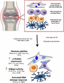
          Cellular senescence is a state of stable proliferation arrest of cells. The senescence pathway has many beneficial effects and is seen to be activated in damaged/stressed cells, as well as during embryonic development and wound healing. However, the persistence and accumulation of senescent cells in various tissues can also impair function and have been implicated in the pathogenesis of many age‐related diseases. Osteoarthritis ( OA), a severely debilitating chronic condition characterized by progressive tissue remodeling and loss of joint function, is the most prevalent disease of the synovial joints, and increasing age is the primary OA risk factor. The profile of inflammatory and catabolic mediators present during the pathogenesis of OA is strikingly similar to the secretory profile observed in ‘classical’ senescent cells. During OA, chondrocytes (the sole cell type present within articular cartilage) exhibit increased levels of various senescence markers, such as senescence‐associated beta‐galactosidase ( SAβGal) activity, telomere attrition, and accumulation of p16ink4a. This suggests the hypothesis that senescence of cells within joint tissues may play a pathological role in the causation of OA. In this review, we discuss the mechanisms by which senescent cells may predispose synovial joints to the development and/or progression of OA, as well as touching upon various epigenetic alterations associated with both OA and senescence.

          Related collections

          Most cited references69

          • Record: found
          • Abstract: found
          • Article: found
          Is Open Access

          The Achilles’ heel of senescent cells: from transcriptome to senolytic drugs

          The healthspan of mice is enhanced by killing senescent cells using a transgenic suicide gene. Achieving the same using small molecules would have a tremendous impact on quality of life and the burden of age-related chronic diseases. Here, we describe the rationale for identification and validation of a new class of drugs termed senolytics, which selectively kill senescent cells. By transcript analysis, we discovered increased expression of pro-survival networks in senescent cells, consistent with their established resistance to apoptosis. Using siRNA to silence expression of key nodes of this network, including ephrins (EFNB1 or 3), PI3Kδ, p21, BCL-xL, or plasminogen-activated inhibitor-2, killed senescent cells, but not proliferating or quiescent, differentiated cells. Drugs targeting these same factors selectively killed senescent cells. Dasatinib eliminated senescent human fat cell progenitors, while quercetin was more effective against senescent human endothelial cells and mouse BM-MSCs. The combination of dasatinib and quercetin was effective in eliminating senescent MEFs. In vivo, this combination reduced senescent cell burden in chronologically aged, radiation-exposed, and progeroid Ercc1 −/Δ mice. In old mice, cardiac function and carotid vascular reactivity were improved 5 days after a single dose. Following irradiation of one limb in mice, a single dose led to improved exercise capacity for at least 7 months following drug treatment. Periodic drug administration extended healthspan in Ercc1 −/Δ mice, delaying age-related symptoms and pathology, osteoporosis, and loss of intervertebral disk proteoglycans. These results demonstrate the feasibility of selectively ablating senescent cells and the efficacy of senolytics for alleviating symptoms of frailty and extending healthspan.
            Bookmark
            • Record: found
            • Abstract: found
            • Article: not found

            Persistent DNA damage signaling triggers senescence-associated inflammatory cytokine secretion

            Cellular senescence suppresses cancer by stably arresting the proliferation of damaged cells1. Paradoxically, senescent cells also secrete factors that alter tissue microenvironments2. The pathways regulating this secretion are unknown. We show that damaged human cells develop persistent chromatin lesions bearing hallmarks of DNA double-strand breaks (DSBs), which initiate increased secretion of inflammatory cytokines such as interleukin-6 (IL-6). Cytokine secretion occurred only after establishment of persistent DNA damage signaling, usually associated with senescence, not after transient DNA damage responses (DDR). Initiation and maintenance of this cytokine response required the DDR proteins ATM, NBS1 and CHK2, but not the cell cycle arrest enforcers p53 and pRb. ATM was also essential for IL-6 secretion during oncogene-induced senescence and by damaged cells that bypass senescence. Further, DDR activity and IL-6 were elevated in human cancers, and ATM-depletion suppressed the ability of senescent cells to stimulate IL-6-dependent cancer cell invasiveness. Thus, in addition to orchestrating cell cycle checkpoints and DNA repair, a novel and important role of the DDR is to allow damaged cells to communicate their compromised state to the surrounding tissue.
              Bookmark
              • Record: found
              • Abstract: found
              • Article: not found

              Oncogenic ras provokes premature cell senescence associated with accumulation of p53 and p16INK4a.

              Oncogenic ras can transform most immortal rodent cells to a tumorigenic state. However, transformation of primary cells by ras requires either a cooperating oncogene or the inactivation of tumor suppressors such as p53 or p16. Here we show that expression of oncogenic ras in primary human or rodent cells results in a permanent G1 arrest. The arrest induced by ras is accompanied by accumulation of p53 and p16, and is phenotypically indistinguishable from cellular senescence. Inactivation of either p53 or p16 prevents ras-induced arrest in rodent cells, and E1A achieves a similar effect in human cells. These observations suggest that the onset of cellular senescence does not simply reflect the accumulation of cell divisions, but can be prematurely activated in response to an oncogenic stimulus. Negation of ras-induced senescence may be relevant during multistep tumorigenesis.
                Bookmark

                Author and article information

                Contributors
                taranjitsingh.rai@uws.ac.uk
                Journal
                Aging Cell
                Aging Cell
                10.1111/(ISSN)1474-9726
                ACEL
                Aging Cell
                John Wiley and Sons Inc. (Hoboken )
                1474-9718
                1474-9726
                26 January 2017
                April 2017
                : 16
                : 2 ( doiID: 10.1111/acel.2017.16.issue-2 )
                : 210-218
                Affiliations
                [ 1 ] Institute of Biomedical and Environmental Health ResearchUniversity of the West of Scotland Paisley PA1 2BEUK
                Author notes
                [*] [* ] Correspondence

                Taranjit Singh Rai, PhD, Institute of Biomedical and Environmental Health Research, University of West of Scotland, Room F247, Paisley PA1 2BE, UK. Tel: +0141 848 3712; fax: +0141 848 3289 ; e‐mail: taranjitsingh.rai@ 123456uws.ac.uk

                Author information
                http://orcid.org/0000-0002-7276-7492
                Article
                ACEL12562
                10.1111/acel.12562
                5334539
                28124466
                12970651-dec9-4767-bf58-2f7b26061da3
                © 2017 The Authors. Aging Cell published by the Anatomical Society and John Wiley & Sons Ltd.

                This is an open access article under the terms of the Creative Commons Attribution License, which permits use, distribution and reproduction in any medium, provided the original work is properly cited.

                History
                : 25 November 2016
                Page count
                Figures: 3, Tables: 0, Pages: 9, Words: 9325
                Categories
                Review
                Reviews
                Custom metadata
                2.0
                acel12562
                April 2017
                Converter:WILEY_ML3GV2_TO_NLMPMC version:5.0.9 mode:remove_FC converted:23.03.2017

                Cell biology
                cellular senescence,epigenetics,osteoarthritis
                Cell biology
                cellular senescence, epigenetics, osteoarthritis

                Comments

                Comment on this article