16
views
0
recommends
+1 Recommend
0 collections
    0
    shares
      • Record: found
      • Abstract: found
      • Article: found
      Is Open Access

      Comprehensive structure and functional adaptations of the yeast nuclear pore complex

      research-article

      Read this article at

      Bookmark
          There is no author summary for this article yet. Authors can add summaries to their articles on ScienceOpen to make them more accessible to a non-specialist audience.

          SUMMARY

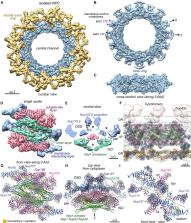
          Nuclear pore complexes (NPCs) mediate the nucleocytoplasmic transport of macromolecules. Here we provide a structure of the isolated yeast NPC in which the inner ring is resolved by cryo-EM at sub-nanometer resolution to show how flexible connectors tie together different structural and functional layers. These connectors may be targets for phosphorylation and regulated disassembly in cells with an open mitosis. Moreover, some nucleoporin pairs and transport factors have similar interaction motifs, which suggests an evolutionary and mechanistic link between assembly and transport. We provide evidence for three major NPC variants that may foreshadow functional specializations at the nuclear periphery. Cryo-electron tomography extended these studies, providing a model of the in situ NPC with a radially expanded inner ring. Our comprehensive model reveals features of the nuclear basket and central transporter, suggests a role for the lumenal Pom152 ring in restricting dilation, and highlights structural plasticity that may be required for transport.

          In brief

          A comprehensive model of the yeast NPC reveals an interconnected architecture of the core scaffold and provides an understanding of the isoforms and structural plasticity that may be associated with different functional states.

          Graphical Abstract

          Related collections

          Most cited references115

          • Record: found
          • Abstract: found
          • Article: found
          Is Open Access

          Highly accurate protein structure prediction with AlphaFold

          Proteins are essential to life, and understanding their structure can facilitate a mechanistic understanding of their function. Through an enormous experimental effort 1 – 4 , the structures of around 100,000 unique proteins have been determined 5 , but this represents a small fraction of the billions of known protein sequences 6 , 7 . Structural coverage is bottlenecked by the months to years of painstaking effort required to determine a single protein structure. Accurate computational approaches are needed to address this gap and to enable large-scale structural bioinformatics. Predicting the three-dimensional structure that a protein will adopt based solely on its amino acid sequence—the structure prediction component of the ‘protein folding problem’ 8 —has been an important open research problem for more than 50 years 9 . Despite recent progress 10 – 14 , existing methods fall far short of atomic accuracy, especially when no homologous structure is available. Here we provide the first computational method that can regularly predict protein structures with atomic accuracy even in cases in which no similar structure is known. We validated an entirely redesigned version of our neural network-based model, AlphaFold, in the challenging 14th Critical Assessment of protein Structure Prediction (CASP14) 15 , demonstrating accuracy competitive with experimental structures in a majority of cases and greatly outperforming other methods. Underpinning the latest version of AlphaFold is a novel machine learning approach that incorporates physical and biological knowledge about protein structure, leveraging multi-sequence alignments, into the design of the deep learning algorithm. AlphaFold predicts protein structures with an accuracy competitive with experimental structures in the majority of cases using a novel deep learning architecture.
            Bookmark
            • Record: found
            • Abstract: found
            • Article: not found

            UCSF Chimera--a visualization system for exploratory research and analysis.

            The design, implementation, and capabilities of an extensible visualization system, UCSF Chimera, are discussed. Chimera is segmented into a core that provides basic services and visualization, and extensions that provide most higher level functionality. This architecture ensures that the extension mechanism satisfies the demands of outside developers who wish to incorporate new features. Two unusual extensions are presented: Multiscale, which adds the ability to visualize large-scale molecular assemblies such as viral coats, and Collaboratory, which allows researchers to share a Chimera session interactively despite being at separate locales. Other extensions include Multalign Viewer, for showing multiple sequence alignments and associated structures; ViewDock, for screening docked ligand orientations; Movie, for replaying molecular dynamics trajectories; and Volume Viewer, for display and analysis of volumetric data. A discussion of the usage of Chimera in real-world situations is given, along with anticipated future directions. Chimera includes full user documentation, is free to academic and nonprofit users, and is available for Microsoft Windows, Linux, Apple Mac OS X, SGI IRIX, and HP Tru64 Unix from http://www.cgl.ucsf.edu/chimera/. Copyright 2004 Wiley Periodicals, Inc.
              Bookmark
              • Record: found
              • Abstract: found
              • Article: not found

              MotionCor2: anisotropic correction of beam-induced motion for improved cryo-electron microscopy

              MotionCor2 software corrects for beam-induced sample motion, improving the resolution of cryo-EM reconstructions.
                Bookmark

                Author and article information

                Journal
                0413066
                2830
                Cell
                Cell
                Cell
                0092-8674
                1097-4172
                22 January 2022
                20 January 2022
                03 January 2022
                17 March 2022
                : 185
                : 2
                : 361-378.e25
                Affiliations
                [1 ]Department of Physiology and Biophysics, Boston University School of Medicine, 700 Albany Street, Boston, MA 02118, USA
                [2 ]Section of Molecular Biology, Division of Biological Sciences, University of California San Diego, La Jolla, CA 92093, USA
                [3 ]Department of Biochemistry and Molecular Pharmacology, University of Massachusetts Medical School, 364 Plantation Street, Worcester, MA 01605, USA
                [4 ]Department of Bioengineering and Therapeutic Sciences, University of California, San Francisco, San Francisco, CA, USA
                [5 ]Laboratory of Cellular and Structural Biology, The Rockefeller University, New York, NY 10065, USA
                [6 ]Department of Cell Biology, University of Pittsburgh, Pittsburgh, PA, USA
                [7 ]Laboratory of Mass Spectrometry and Gaseous Ion Chemistry, The Rockefeller University, New York, NY, USA
                [8 ]School of Physics, Georgia Institute of Technology, Atlanta, GA 30332, USA
                [9 ]Stowers Institute for Medical Research, Kansas City, MO, USA
                [10 ]Department of Molecular and Integrative Physiology, University of Kansas Medical Center, Kansas City, KS, USA
                [11 ]Verna and Marrs McLean Department of Biochemistry and Molecular Biology, Baylor College of Medicine, 1 Baylor Plaza, Houston, Texas 77030, USA
                [12 ]Department of Cellular and Molecular Pharmacology, San Francisco, San Francisco, CA 94158, USA
                [13 ]Quantitative Biosciences Institute, University of California San Francisco, San Francisco, CA 94158, USA
                [14 ]Department of Pharmaceutical Chemistry, University of California San Francisco, San Francisco, CA 94158, USA
                [15 ]Howard Hughes Medical Institute, University of California, San Diego, La Jolla, CA 92093, USA
                [16 ]Present address: Ikerbasque, Basque Foundation for Science, 48013 Bilbao, Spain
                [17 ]Present address: Instituto Biofisika (UPV/EHU, CSIC), University of the Basque Country, 48940 Leioa, Spain
                [18 ]These authors contributed equally
                [19 ]Lead contact
                Author notes

                AUTHOR CONTRIBUTIONS

                Conceptualization, C.W.A., S.J.L., E.V., S.L.J., J.F.-M., and M.P.R.; Investigation, C.W.A., J.F.-M., I.N., D.S., S.S., K.S., C.X., F.F., Y.S., J.W., and Z.Y.; Formal Analysis, C.W.A., C.O., D.S., S.J.L., I.E., J.C.G., J.W., and J.U.; Writing, C.W.A., J.F.-M., D.S., J.M.V., S.L.J., S.J.L., A.S., B.T.C., I.E., E.V., and M.P.R.; Funding Acquisition, C.W.A., E.V., S.J.L., A.S., S.L.J., B.T.C., and M.P.R.; Supervision, C.W.A., E.V., S.J.L., S.L.J., A.S., B.T.C., and M.P.R.

                Article
                NIHMS1767905
                10.1016/j.cell.2021.12.015
                8928745
                34982960
                11d845e3-9a0f-47d9-989d-42f1f3cbcc65

                This is an open access article under the CC BY-NC-ND license ( http://creativecommons.org/licenses/by-nc-nd/4.0/).

                History
                Categories
                Article

                Cell biology
                Cell biology

                Comments

                Comment on this article