26
views
0
recommends
+1 Recommend
0 collections
    0
    shares
      • Record: found
      • Abstract: found
      • Article: found
      Is Open Access

      Early long-term administration of the CSF1R inhibitor PLX3397 ablates microglia and reduces accumulation of intraneuronal amyloid, neuritic plaque deposition and pre-fibrillar oligomers in 5XFAD mouse model of Alzheimer’s disease

      research-article

      Read this article at

      Bookmark
          There is no author summary for this article yet. Authors can add summaries to their articles on ScienceOpen to make them more accessible to a non-specialist audience.

          Abstract

          Background

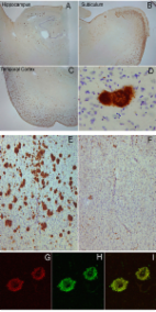
          Besides the two main classical features of amyloid beta aggregation and tau-containing neurofibrillary tangle deposition, neuroinflammation plays an important yet unclear role in the pathophysiology of Alzheimer’s disease (AD). Microglia are believed to be key mediators of neuroinflammation during AD and responsible for the regulation of brain homeostasis by balancing neurotoxicity and neuroprotective events. We have previously reported evidence that neuritic plaques are derived from dead neurons that have accumulated intraneuronal amyloid and further recruit Iba1-positive cells, which play a role in either neuronal demise or neuritic plaque maturation or both.

          Methods

          To study the impact of microglia on neuritic plaque development, we treated two-month-old 5XFAD mice with a selective colony stimulation factor 1 receptor (CSF1R) inhibitor, PLX3397, for a period of 3 months, resulting in a significant ablation of microglia. Directly after this treatment, we analyzed the amount of intraneuronal amyloid and neuritic plaques and performed behavioral studies including Y-maze, fear conditioning and elevated plus maze.

          Results

          We found that early long-term PLX3397 administration results in a dramatic reduction of both intraneuronal amyloid as well as neuritic plaque deposition. PLX3397 treated young 5XFAD mice also displayed a significant decrease of soluble fibrillar amyloid oligomers in brain lysates, a depletion of soluble pre-fibrillar oligomers in plasma and an improvement in cognitive function measured by fear conditioning tests.

          Conclusions

          Our findings demonstrate that CSF1R signaling, either directly on neurons or mediated by microglia, is crucial for the accumulation of intraneuronal amyloid and formation of neuritic plaques, suggesting that these two events are serially linked in a causal pathway leading to neurodegeneration and neuritic plaque formation. CSF1R inhibitors represent potential preventative or therapeutic approach that target the very earliest stages of the formation of intraneuronal amyloid and neuritic plaques.

          Related collections

          Most cited references22

          • Record: found
          • Abstract: found
          • Article: not found

          Common structure of soluble amyloid oligomers implies common mechanism of pathogenesis.

          Soluble oligomers are common to most amyloids and may represent the primary toxic species of amyloids, like the Abeta peptide in Alzheimer's disease (AD). Here we show that all of the soluble oligomers tested display a common conformation-dependent structure that is unique to soluble oligomers regardless of sequence. The in vitro toxicity of soluble oligomers is inhibited by oligomer-specific antibody. Soluble oligomers have a unique distribution in human AD brain that is distinct from fibrillar amyloid. These results indicate that different types of soluble amyloid oligomers have a common structure and suggest they share a common mechanism of toxicity.
            Bookmark
            • Record: found
            • Abstract: found
            • Article: found
            Is Open Access

            Fibril specific, conformation dependent antibodies recognize a generic epitope common to amyloid fibrils and fibrillar oligomers that is absent in prefibrillar oligomers

            Background Amyloid-related degenerative diseases are associated with the accumulation of misfolded proteins as amyloid fibrils in tissue. In Alzheimer disease (AD), amyloid accumulates in several distinct types of insoluble plaque deposits, intracellular Aβ and as soluble oligomers and the relationships between these deposits and their pathological significance remains unclear. Conformation dependent antibodies have been reported that specifically recognize distinct assembly states of amyloids, including prefibrillar oligomers and fibrils. Results We immunized rabbits with a morphologically homogeneous population of Aβ42 fibrils. The resulting immune serum (OC) specifically recognizes fibrils, but not random coil monomer or prefibrillar oligomers, indicating fibrils display a distinct conformation dependent epitope that is absent in prefibrillar oligomers. The fibril epitope is also displayed by fibrils of other types of amyloids, indicating that the epitope is a generic feature of the polypeptide backbone. The fibril specific antibody also recognizes 100,000 × G soluble fibrillar oligomers ranging in size from dimer to greater than 250 kDa on western blots. The fibrillar oligomers recognized by OC are immunologically distinct from prefibrillar oligomers recognized by A11, even though their sizes overlap broadly, indicating that size is not a reliable indicator of oligomer conformation. The immune response to prefibrillar oligomers and fibrils is not sequence specific and antisera of the same specificity are produced in response to immunization with islet amyloid polypeptide prefibrillar oligomer mimics and fibrils. The fibril specific antibodies stain all types of amyloid deposits in human AD brain. Diffuse amyloid deposits stain intensely with anti-fibril antibody although they are thioflavin S negative, suggesting that they are indeed fibrillar in conformation. OC also stains islet amyloid deposits in transgenic mouse models of type II diabetes, demonstrating its generic specificity for amyloid fibrils. Conclusion Since the fibril specific antibodies are conformation dependent, sequence-independent, and recognize epitopes that are distinct from those present in prefibrillar oligomers, they may have broad utility for detecting and characterizing the accumulation of amyloid fibrils and fibrillar type oligomers in degenerative diseases.
              Bookmark
              • Record: found
              • Abstract: found
              • Article: found
              Is Open Access

              Neuron loss in the 5XFAD mouse model of Alzheimer’s disease correlates with intraneuronal Aβ42 accumulation and Caspase-3 activation

              Background Although the mechanism of neuron loss in Alzheimer’s disease (AD) is enigmatic, it is associated with cerebral accumulation of Aβ42. The 5XFAD mouse model of amyloid deposition expresses five familial AD (FAD) mutations that are additive in driving Aβ42 overproduction. 5XFAD mice exhibit intraneuronal Aβ42 accumulation at 1.5 months, amyloid deposition at 2 months, and memory deficits by 4 months of age. Results Here, we demonstrate by unbiased stereology that statistically significant neuron loss occurs by 9 months of age in 5XFAD mice. We validated two Aβ42-selective antibodies by immunostaining 5XFAD; BACE1−/− bigenic brain sections and then used these antibodies to show that intraneuronal Aβ42 and amyloid deposition develop in the same regions where neuron loss is observed in 5XFAD brain. In 5XFAD neuronal soma, intraneuronal Aβ42 accumulates in puncta that co-label for Transferrin receptor and LAMP-1, indicating endosomal and lysosomal localization, respectively. In addition, in young 5XFAD brains, we observed activated Caspase-3 in the soma and proximal dendrites of intraneuronal Aβ42-labeled neurons. In older 5XFAD brains, we found activated Caspase-3-positive punctate accumulations that co-localize with the neuronal marker class III β-tubulin, suggesting neuron loss by apoptosis. Conclusions Together, our results indicate a temporal sequence of intraneuronal Aβ42 accumulation, Caspase-3 activation, and neuron loss that implies a potential apoptotic mechanism of neuron death in the 5XFAD mouse.
                Bookmark

                Author and article information

                Contributors
                cglabe@uci.edu
                Journal
                Mol Neurodegener
                Mol Neurodegener
                Molecular Neurodegeneration
                BioMed Central (London )
                1750-1326
                1 March 2018
                1 March 2018
                2018
                : 13
                : 11
                Affiliations
                [1 ]ISNI 0000 0001 0668 7243, GRID grid.266093.8, Department of Molecular Biology and Biochemistry, , University of California, ; Irvine, USA
                [2 ]ISNI 0000 0001 0619 1117, GRID grid.412125.1, Biochemistry Department, Faculty of Science and Experimental Biochemistry Unit, King Fahd Medical Research Center, , King Abdulaziz University, ; Jeddah, 21589 Saudi Arabia
                [3 ]ISNI 0000 0001 0668 7243, GRID grid.266093.8, Institute for Memory Impairments and Neurological Disorders (UCIMIND), , University of California, ; Irvine, USA
                Article
                244
                10.1186/s13024-018-0244-x
                5831225
                29490706
                11cf77a8-9616-41b3-9bd4-58cd03c28cda
                © The Author(s). 2018

                Open AccessThis article is distributed under the terms of the Creative Commons Attribution 4.0 International License ( http://creativecommons.org/licenses/by/4.0/), which permits unrestricted use, distribution, and reproduction in any medium, provided you give appropriate credit to the original author(s) and the source, provide a link to the Creative Commons license, and indicate if changes were made. The Creative Commons Public Domain Dedication waiver ( http://creativecommons.org/publicdomain/zero/1.0/) applies to the data made available in this article, unless otherwise stated.

                History
                : 21 December 2017
                : 22 February 2018
                Funding
                Funded by: FundRef http://dx.doi.org/10.13039/100000049, National Institute on Aging;
                Award ID: AG00538
                Award ID: AG00538
                Award ID: AG056507
                Award ID: AG027544
                Award Recipient :
                Funded by: FundRef http://dx.doi.org/10.13039/100000957, Alzheimer's Association;
                Award ID: NIRG-15-363477
                Award Recipient :
                Funded by: FundRef http://dx.doi.org/10.13039/100007625, Cure Alzheimer's Fund;
                Categories
                Research Article
                Custom metadata
                © The Author(s) 2018

                Neurosciences
                alzheimer’s disease,amyloid beta,plaques,intraneuronal amyloid,microglia,neuroinflammation

                Comments

                Comment on this article