81
views
0
recommends
+1 Recommend
0 collections
    0
    shares
      • Record: found
      • Abstract: found
      • Article: found
      Is Open Access

      Autophagic degradation of dBruce controls DNA fragmentation in nurse cells during late Drosophila melanogaster oogenesis

      research-article

      Read this article at

      Bookmark
          There is no author summary for this article yet. Authors can add summaries to their articles on ScienceOpen to make them more accessible to a non-specialist audience.

          Abstract

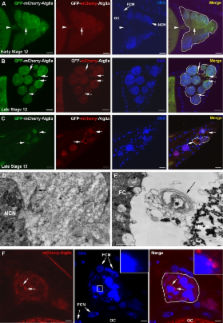
          Blocking autophagy protects the apoptosis inhibitor dBruce from destruction and promotes nurse cell survival in developing egg chambers.

          Abstract

          Autophagy is an evolutionarily conserved pathway responsible for degradation of cytoplasmic material via the lysosome. Although autophagy has been reported to contribute to cell death, the underlying mechanisms remain largely unknown. In this study, we show that autophagy controls DNA fragmentation during late oogenesis in Drosophila melanogaster. Inhibition of autophagy by genetically removing the function of the autophagy genes atg1, atg13, and vps34 resulted in late stage egg chambers that contained persisting nurse cell nuclei without fragmented DNA and attenuation of caspase-3 cleavage. The Drosophila inhibitor of apoptosis (IAP) dBruce was found to colocalize with the autophagic marker GFP-Atg8a and accumulated in autophagy mutants. Nurse cells lacking Atg1 or Vps34 in addition to dBruce contained persisting nurse cell nuclei with fragmented DNA. This indicates that autophagic degradation of dBruce controls DNA fragmentation in nurse cells. Our results reveal autophagic degradation of an IAP as a novel mechanism of triggering cell death and thereby provide a mechanistic link between autophagy and cell death.

          Related collections

          Most cited references34

          • Record: found
          • Abstract: found
          • Article: not found

          Role and regulation of starvation-induced autophagy in the Drosophila fat body.

          In response to starvation, eukaryotic cells recover nutrients through autophagy, a lysosomal-mediated process of cytoplasmic degradation. Autophagy is known to be inhibited by TOR signaling, but the mechanisms of autophagy regulation and its role in TOR-mediated cell growth are unclear. Here, we show that signaling through TOR and its upstream regulators PI3K and Rheb is necessary and sufficient to suppress starvation-induced autophagy in the Drosophila fat body. In contrast, TOR's downstream effector S6K promotes rather than suppresses autophagy, suggesting S6K downregulation may limit autophagy during extended starvation. Despite the catabolic potential of autophagy, disruption of conserved components of the autophagic machinery, including ATG1 and ATG5, does not restore growth to TOR mutant cells. Instead, inhibition of autophagy enhances TOR mutant phenotypes, including reduced cell size, growth rate, and survival. Thus, in cells lacking TOR, autophagy plays a protective role that is dominant over its potential role as a growth suppressor.
            Bookmark
            • Record: found
            • Abstract: found
            • Article: not found

            Ref(2)P, the Drosophila melanogaster homologue of mammalian p62, is required for the formation of protein aggregates in adult brain

            p62 has been proposed to mark ubiquitinated protein bodies for autophagic degradation. We report that the Drosophila melanogaster p62 orthologue, Ref(2)P, is a regulator of protein aggregation in the adult brain. We demonstrate that Ref(2)P localizes to age-induced protein aggregates as well as to aggregates caused by reduced autophagic or proteasomal activity. A similar localization to protein aggregates is also observed in D. melanogaster models of human neurodegenerative diseases. Although atg8a autophagy mutant flies show accumulation of ubiquitin- and Ref(2)P-positive protein aggregates, this is abrogated in atg8a/ref(2)P double mutants. Both the multimerization and ubiquitin binding domains of Ref(2)P are required for aggregate formation in vivo. Our findings reveal a major role for Ref(2)P in the formation of ubiquitin-positive protein aggregates both under physiological conditions and when normal protein turnover is inhibited.
              Bookmark
              • Record: found
              • Abstract: found
              • Article: not found

              Growth arrest and autophagy are required for salivary gland cell degradation in Drosophila.

              Autophagy is a catabolic process that is negatively regulated by growth and has been implicated in cell death. We find that autophagy is induced following growth arrest and precedes developmental autophagic cell death of Drosophila salivary glands. Maintaining growth by expression of either activated Ras or positive regulators of the class I phosphoinositide 3-kinase (PI3K) pathway inhibits autophagy and blocks salivary gland cell degradation. Developmental degradation of salivary glands is also inhibited in autophagy gene (atg) mutants. Caspases are active in PI3K-expressing and atg mutant salivary glands, and combined inhibition of both autophagy and caspases increases suppression of gland degradation. Further, induction of autophagy is sufficient to induce premature cell death in a caspase-independent manner. Our results provide in vivo evidence that growth arrest, autophagy, and atg genes are required for physiological autophagic cell death and that multiple degradation pathways cooperate in the efficient clearance of cells during development.
                Bookmark

                Author and article information

                Journal
                J Cell Biol
                J. Cell Biol
                jcb
                The Journal of Cell Biology
                The Rockefeller University Press
                0021-9525
                1540-8140
                23 August 2010
                : 190
                : 4
                : 523-531
                Affiliations
                [1 ]Centre for Cancer Biomedicine and [2 ]Department of Biochemistry, Institute for Cancer Research, The Norwegian Radium Hospital, Oslo University Hospital, N-0310, Oslo, Norway
                [3 ]Department of Cancer Biology, University of Massachusetts Medical School, Worcester, MA 01605
                [4 ]Molecular Cancer Research Group, Institute of Medical Biology, University of Tromsø, 9037 Tromsø, Norway
                [5 ]Department of Technology, Sør-Trøndelag University College, 7004 Trondheim, Norway
                Author notes
                Correspondence to Ioannis P. Nezis: Ioannis.Nezis@ 123456rr-research.no ; or Harald Stenmark: stenmark@ 123456ulrik.uio.no
                Article
                201002035
                10.1083/jcb.201002035
                2928014
                20713604
                11c1b385-d00d-4e11-9e26-18120c777792
                © 2010 Nezis et al.

                This article is distributed under the terms of an Attribution–Noncommercial–Share Alike–No Mirror Sites license for the first six months after the publication date (see http://www.rupress.org/terms). After six months it is available under a Creative Commons License (Attribution–Noncommercial–Share Alike 3.0 Unported license, as described at http://creativecommons.org/licenses/by-nc-sa/3.0/).

                History
                : 8 February 2010
                : 20 July 2010
                Categories
                Research Articles
                Report

                Cell biology
                Cell biology

                Comments

                Comment on this article