8
views
0
recommends
+1 Recommend
0 collections
    0
    shares
      • Record: found
      • Abstract: found
      • Article: found
      Is Open Access

      How Do Cells of the Oligodendrocyte Lineage Affect Neuronal Circuits to Influence Motor Function, Memory and Mood?

      review-article

      Read this article at
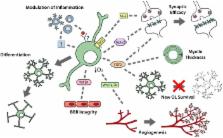
      Bookmark
          There is no author summary for this article yet. Authors can add summaries to their articles on ScienceOpen to make them more accessible to a non-specialist audience.

          Abstract

          Oligodendrocyte progenitor cells (OPCs) are immature cells in the central nervous system (CNS) that can rapidly respond to changes within their environment by modulating their proliferation, motility and differentiation. OPCs differentiate into myelinating oligodendrocytes throughout life, and both cell types have been implicated in maintaining and modulating neuronal function to affect motor performance, cognition and emotional state. However, questions remain about the mechanisms employed by OPCs and oligodendrocytes to regulate circuit function, including whether OPCs can only influence circuits through their generation of new oligodendrocytes, or can play other regulatory roles within the CNS. In this review, we detail the molecular and cellular mechanisms that allow OPCs, newborn oligodendrocytes and pre-existing oligodendrocytes to regulate circuit function and ultimately influence behavioral outcomes.

          Related collections

          Most cited references97

          • Record: found
          • Abstract: found
          • Article: not found

          Competing waves of oligodendrocytes in the forebrain and postnatal elimination of an embryonic lineage.

          The developmental origin of oligodendrocyte progenitors (OLPs) in the forebrain has been controversial. We now show, by Cre-lox fate mapping in transgenic mice, that the first OLPs originate in the medial ganglionic eminence (MGE) and anterior entopeduncular area (AEP) in the ventral forebrain. From there, they populate the entire embryonic telencephalon including the cerebral cortex before being joined by a second wave of OLPs from the lateral and/or caudal ganglionic eminences (LGE and CGE). Finally, a third wave arises within the postnatal cortex. When any one population is destroyed at source by the targeted expression of diphtheria toxin, the remaining cells take over and the mice survive and behave normally, with a normal complement of oligodendrocytes and myelin. Thus, functionally redundant populations of OLPs compete for space in the developing brain. Notably, the embryonic MGE- and AEP-derived population is eliminated during postnatal life, raising questions about the nature and purpose of the competition.
            Bookmark
            • Record: found
            • Abstract: found
            • Article: not found

            Disruption of Cnp1 uncouples oligodendroglial functions in axonal support and myelination.

            Myelination of axons by oligodendrocytes enables rapid impulse propagation in the central nervous system. But long-term interactions between axons and their myelin sheaths are poorly understood. Here we show that Cnp1, which encodes 2',3'-cyclic nucleotide phosphodiesterase in oligodendrocytes, is essential for axonal survival but not for myelin assembly. In the absence of glial cyclic nucleotide phosphodiesterase, mice developed axonal swellings and neurodegeneration throughout the brain, leading to hydrocephalus and premature death. But, in contrast to previously studied myelin mutants, the ultrastructure, periodicity and physical stability of myelin were not altered in these mice. Genetically, the chief function of glia in supporting axonal integrity can thus be completely uncoupled from its function in maintaining compact myelin. Oligodendrocyte dysfunction, such as that in multiple sclerosis lesions, may suffice to cause secondary axonal loss.
              Bookmark
              • Record: found
              • Abstract: found
              • Article: not found

              NG2 cells generate both oligodendrocytes and gray matter astrocytes.

              NG2 glia constitute a fourth major glial cell type in the mammalian central nervous system (CNS) that is distinct from other cell types. Although circumstantial evidence suggests that some NG2 glia differentiate into oligodendrocytes, their in vivo fate has not been directly examined. We have used the bacterial artificial chromosome (BAC) modification technique to generate transgenic mice that express DsRed or Cre specifically in NG2-expressing (NG2+) cells. In NG2DsRedBAC transgenic mice, DsRed was expressed specifically in NG2+ cells throughout the postnatal CNS. When the differentiation potential of NG2+ cells in vitro was examined using DsRed+NG2+ cells purified from perinatal transgenic brains, the majority of the cells either remained as NG2+ cells or differentiated into oligodendrocytes. In addition, DsRed+NG2+ cells also differentiated into astrocytes. The in vivo fate of NG2 glia was examined in mice that were double transgenic for NG2creBAC and the Cre reporter Z/EG. In the double transgenic mice, the Cre reporter EGFP was detected in myelinating oligodendrocytes and in a subpopulation of protoplasmic astrocytes in the gray matter of ventrolateral forebrain but not in fibrous astrocytes of white matter. These observations suggest that NG2+ cells are precursors of oligodendrocytes and some protoplasmic astrocytes in gray matter.
                Bookmark

                Author and article information

                Contributors
                Journal
                Front Cell Neurosci
                Front Cell Neurosci
                Front. Cell. Neurosci.
                Frontiers in Cellular Neuroscience
                Frontiers Media S.A.
                1662-5102
                16 November 2018
                2018
                : 12
                : 399
                Affiliations
                Menzies Institute for Medical Research, University of Tasmania , Hobart, TAS, Australia
                Author notes

                Edited by: Philippe Isope, Centre National de la Recherche Scientifique (CNRS), France

                Reviewed by: Fernando de Castro, Instituto Cajal (IC), Spain; Herbert Hildebrandt, Hannover Medical School, Germany

                *Correspondence: Kaylene M. Young kaylene.young@ 123456utas.edu.au

                These authors have contributed equally to this work

                Article
                10.3389/fncel.2018.00399
                6262292
                30524235
                109d92d0-a8bf-4ea6-b281-507b77c14382
                Copyright © 2018 Pepper, Pitman, Cullen and Young.

                This is an open-access article distributed under the terms of the Creative Commons Attribution License (CC BY). The use, distribution or reproduction in other forums is permitted, provided the original author(s) and the copyright owner(s) are credited and that the original publication in this journal is cited, in accordance with accepted academic practice. No use, distribution or reproduction is permitted which does not comply with these terms.

                History
                : 30 July 2018
                : 17 October 2018
                Page count
                Figures: 3, Tables: 0, Equations: 0, References: 156, Pages: 14, Words: 11908
                Categories
                Neuroscience
                Review

                Neurosciences
                ng2 glia,myelin,oligodendrocyte,anxiety,motor function,depression,learning,neuronal activity
                Neurosciences
                ng2 glia, myelin, oligodendrocyte, anxiety, motor function, depression, learning, neuronal activity

                Comments

                Comment on this article