1
views
0
recommends
+1 Recommend
0 collections
    0
    shares
      • Record: found
      • Abstract: found
      • Article: found
      Is Open Access

      Differential Expression Patterns of Rspondin Family and Leucine-Rich Repeat-Containing G-Protein Coupled Receptors in Chondrocytes and Osteoblasts

      research-article
      , M.Sc., , Ph.D., , Ph.D., , M.D., Ph.D. * , , Ph.D. *
      Cell Journal (Yakhteh)
      Royan Institute
      Chondrocyte, LGR, Osteoarthritis, Osteoblast, R-spondin

      Read this article at

      Bookmark
          There is no author summary for this article yet. Authors can add summaries to their articles on ScienceOpen to make them more accessible to a non-specialist audience.

          Abstract

          Objective

          Rspondins (RSPOs) are regarded as the significant modulators of WNT signaling pathway and they are expressed dynamically during developmental stages. Since in osteoarthritis (OA) both cartilage and subchondral bone suffer damages and WNT signaling pathway has a crucial role in their maintenance, the objective of the study was to analyze expression profile of RSPO family and its receptors [leucine-rich repeat-containing G-protein coupled receptors (LGRs)] in OA tissue samples as well as in differentiating chondrocytes and osteoblasts.

          Materials and Methods:

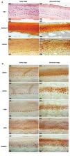
          In this experimental study, human early and advanced stage of OA tissue samples were analyzed for the morphological changes of articular cartilage by hematoxylin and eosin (H&E) staining, safranin-O staining and lubricin immunostaining. RSPOs and LGRs expression were confirmed by immunohistochemistry. Human primary chondrocytes and human osteoblast cell line, SaOS-2, were cultured in differentiation medium till day 14 and they were analyzed in terms of expression of RSPOs, LGRs and specific marker for chondrogenesis and osteogenesis by western blotting and quantitative reverse transcription polymerase chain reaction (qRT-PCR).

          Results

          Advanced stage OA tissue samples showed increased expression of RSPO1 and LGR6 in a region close to subchondral bone. While RSPO2 and LGR5 expression were seen overlapping in the deep region of articular cartilage. Differentiating chondrocytes demonstrated elevated expression of RSPO2 and LGR5 from day 7 to day 14, whereas, osteoblasts undergoing differentiation showed enhanced expression of RSPO1 and LGR6 from day 2 to day 14. Under tumor necrosis factor alpha (TNFα) stimulatory conditions, RSPO2 and RSPO1 recovered the suppressed expression of inflammatory, chondrogenic and osteogenic markers, respectively. A recovery in the stability of β-catenin was also noticed in both cases.

          Conclusion:

          Spatial expression of RSPOs during progression of OA might be dynamically controlled by cartilage and subchondral bone. Interplay amid chondrocytes and osteoblasts, via RSPOs, might provide probable mechanisms for treating inflammatory pathogenic conditions like OA.

          Related collections

          Most cited references40

          • Record: found
          • Abstract: found
          • Article: not found

          The bone-cartilage unit in osteoarthritis.

          Osteoarthritis (OA) refers to a group of mechanically-induced joint disorders to which both genetic and acquired factors contribute. Current pathophysiological concepts focus on OA as a disease of the whole joint. Within these models, the functional unit formed by the articular cartilage and the subchondral bone seems to be of particular interest. Cartilage and bone receive and dissipate the stress associated with movement and loading, and are therefore continuously challenged biomechanically. Recent data support the view that cartilage and bone can communicate over the calcified tissue barrier; vessels reach out from bone into the cartilage zone, patches of uncalcified cartilage are in contact with bone, and microcracks and fissures further facilitate transfer of molecules. Several molecular signaling pathways such as bone morphogenetic proteins and Wnts are hypothesized to have a role in OA and can activate cellular and molecular processes in both cartilage and bone cells. In addition, intracellular activation of different kinase cascades seems to be involved in the molecular crosstalk between cartilage and bone cells. Further research is required to integrate these different elements into a comprehensive approach that will increase our understanding of the disease processes in OA, and that could lead to the development of specific therapeutics or treatment strategies.
            Bookmark
            • Record: found
            • Abstract: found
            • Article: not found

            Characterization of articular cartilage and subchondral bone changes in the rat anterior cruciate ligament transection and meniscectomized models of osteoarthritis.

            Osteoarthritis (OA) is a chronic joint disease characterized by cartilage destruction, subchondral bone sclerosis, and osteophyte formation. Subchondral bone stiffness has been proposed to initiate and/or contribute to cartilage deterioration in OA. The purpose of this study was to characterize subchondral bone remodeling, cartilage damage, and osteophytosis during the disease progression in two models of surgically induced OA. Rat knee joints were subjected either to anterior cruciate ligament transection (ACLT) alone or in combination with resection of medial menisci (ACLT + MMx). Histopathological changes in the surgical joints were compared with sham at 1, 2, 4, 6, and 10 weeks post-surgery. Using a modified Mankin scoring system, we demonstrate that articular cartilage damage occurs within 2 weeks post-surgery in both surgical models. Detectable cartilage surface damage and proteoglycan loss were observed as early as 1 week post-surgery. These were followed by the increases in vascular invasion into cartilage, in loss of chondrocyte number and in cell clustering. Histomorphometric analysis revealed subchondral bone loss in both models within 2 weeks post-surgery followed by significant increases in subchondral bone volume relative to sham up to 10 weeks post-surgery. Incidence of osteophyte formation was optimally observed in ACLT joints at 10 weeks and in ACLT + MMx joints at 6 weeks post-surgery. In summary, the two surgically induced rat OA models share many characteristics seen in human and other animal models of OA, including progressive articular cartilage degradation, subchondral bone sclerosis, and osteophyte formation. Moreover, increased subchondral bone resorption is associated with early development of cartilage lesions, which precedes significant cartilage thinning and subchondral bone sclerosis. Together, these findings support a role for bone remodeling in OA pathogenesis and suggest that these rat models are suitable for evaluating bone resorption inhibitors as potential disease-modifying pharmaco-therapies.
              Bookmark
              • Record: found
              • Abstract: found
              • Article: not found

              Activation of beta-catenin signaling in articular chondrocytes leads to osteoarthritis-like phenotype in adult beta-catenin conditional activation mice.

              Osteoarthritis (OA) is a degenerative joint disease, and the mechanism of its pathogenesis is poorly understood. Recent human genetic association studies showed that mutations in the Frzb gene predispose patients to OA, suggesting that the Wnt/beta-catenin signaling may be the key pathway to the development of OA. However, direct genetic evidence for beta-catenin in this disease has not been reported. Because tissue-specific activation of the beta-catenin gene (targeted by Col2a1-Cre) is embryonic lethal, we specifically activated the beta-catenin gene in articular chondrocytes in adult mice by generating beta-catenin conditional activation (cAct) mice through breeding of beta-catenin(fx(Ex3)/fx(Ex3)) mice with Col2a1-CreER(T2) transgenic mice. Deletion of exon 3 of the beta-catenin gene results in the production of a stabilized fusion beta-catenin protein that is resistant to phosphorylation by GSK-3beta. In this study, tamoxifen was administered to the 3- and 6-mo-old Col2a1-CreER(T2);beta-catenin(fx(Ex3)/wt) mice, and tissues were harvested for histologic analysis 2 mo after tamoxifen induction. Overexpression of beta-catenin protein was detected by immunostaining in articular cartilage tissues of beta-catenin cAct mice. In 5-mo-old beta-catenin cAct mice, reduction of Safranin O and Alcian blue staining in articular cartilage tissue and reduced articular cartilage area were observed. In 8-mo-old beta-catenin cAct mice, cell cloning, surface fibrillation, vertical clefting, and chondrophyte/osteophyte formation were observed. Complete loss of articular cartilage layers and the formation of new woven bone in the subchondral bone area were also found in beta-catenin cAct mice. Expression of chondrocyte marker genes, such as aggrecan, Mmp-9, Mmp-13, Alp, Oc, and colX, was significantly increased (3- to 6-fold) in articular chondrocytes derived from beta-catenin cAct mice. Bmp2 but not Bmp4 expression was also significantly upregulated (6-fold increase) in these cells. In addition, we also observed overexpression of beta-catenin protein in the knee joint samples from patients with OA. These findings indicate that activation of beta-catenin signaling in articular chondrocytes in adult mice leads to the premature chondrocyte differentiation and the development of an OA-like phenotype. This study provides direct and definitive evidence about the role of beta-catenin in the development of OA.
                Bookmark

                Author and article information

                Journal
                Cell J
                Cell J
                Royan Institute
                Cell Journal (Yakhteh)
                Royan Institute
                2228-5806
                2228-5814
                Jan-Mar 2021
                22 April 2020
                : 22
                : 4
                : 437-449
                Affiliations
                Institute for Skeletal Aging and Orthopedic Surgery, Hallym University-Chuncheon Sacred Heart Hospital, Chuncheon, Korea
                Author notes
                [*Corresponding Address: ]Institute for Skeletal Aging and Orthopedic Surgery Hallym University-Chuncheon Sacred Heart Hospital ChuncheonKorea Emails: 123sslee@ 123456gmail.com , jsnam88@ 123456hallym.ac.kr
                [#]

                The first two authors equally contributed to this work.

                Article
                Cell-J-22-437
                10.22074/cellj.2021.6927
                7211279
                32347037
                1076eae3-fc05-47ca-8ce8-fb80f982f47e
                Any use, distribution, reproduction or abstract of this publication in any medium, with the exception of commercial purposes, is permitted provided the original work is properly cited.

                This is an open-access article distributed under the terms of the Creative Commons Attribution License, which permits unrestricted use, distribution, and reproduction in any medium, provided the original work is properly cited.

                History
                : 8 May 2019
                : 5 August 2019
                Categories
                Original Article
                Genetics
                Differentiate
                Custom metadata
                Lee YH, Sharma AR, Jagga S, Lee SS, Nam JS. Differential expression patterns of rspondin family and leucine-rich repeat-containing G-protein coupled receptors in chondrocytes and osteoblasts. Cell J. 2021; 22(4): 437-449. doi: 10.22074/cellj.2021.6927. This open-access article has been published under the terms of the Creative Commons Attribution Non-Commercial 3.0 (CC BY-NC 3.0).

                chondrocyte,lgr,osteoarthritis,osteoblast,r-spondin
                chondrocyte, lgr, osteoarthritis, osteoblast, r-spondin

                Comments

                Comment on this article