33
views
0
recommends
+1 Recommend
0 collections
    0
    shares
      • Record: found
      • Abstract: found
      • Article: found
      Is Open Access

      Biomolecular environment, quantification, and intracellular interaction of multifunctional magnetic SERS nanoprobes†

      research-article

      Read this article at

      Bookmark
          There is no author summary for this article yet. Authors can add summaries to their articles on ScienceOpen to make them more accessible to a non-specialist audience.

          Abstract

          Multifunctional composite nanoprobes, Ag–Magnetite and Au–Magnetite, were manipulated in fibroblast cells and characterized using SERS, LA-ICP-MS, and nanotomography.

          Abstract

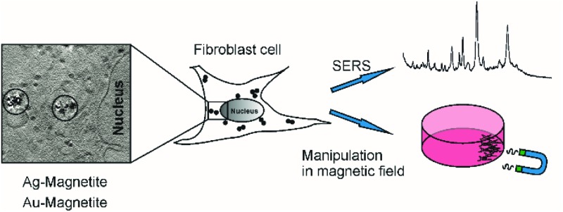
          Multifunctional composite nanoprobes consisting of iron oxide nanoparticles linked to silver and gold nanoparticles, Ag–Magnetite and Au–Magnetite, respectively, were introduced by endocytic uptake into cultured fibroblast cells. The cells containing the non-toxic nanoprobes were shown to be displaceable in an external magnetic field and can be manipulated in microfluidic channels. The distribution of the composite nanostructures that are contained in the endosomal system is discussed on the basis of surface-enhanced Raman scattering (SERS) mapping, quantitative laser ablation inductively coupled plasma mass spectrometry (LA-ICP-MS) micromapping, and cryo soft X-ray tomography (cryo soft-XRT). Cryo soft-XRT of intact, vitrified cells reveals that the composite nanoprobes form intra-endosomal aggregates. The nanoprobes provide SERS signals from the biomolecular composition of their surface in the endosomal environment. The SERS data indicate the high stability of the nanoprobes and of their plasmonic properties in the harsh environment of endosomes and lysosomes. The spectra point at the molecular composition at the surface of the Ag–Magnetite and Au–Magnetite nanostructures that is very similar to that of other composite structures, but different from the composition of pure silver and gold SERS nanoprobes used for intracellular investigations. As shown by the LA-ICP-MS data, the uptake efficiency of the magnetite composites is approximately two to three times higher than that of the pure gold and silver nanoparticles.

          Related collections

          Most cited references71

          • Record: found
          • Abstract: found
          • Article: not found

          Nuclear pore complex is able to transport macromolecules with diameters of about 39 nm.

          Bidirectional transport of macromolecules between the nucleus and the cytoplasm occurs through the nuclear pore complexes (NPCs) by a signal-mediated mechanism that is directed by targeting signals (NLSs) residing on the transported molecules or "cargoes." Nuclear transport starts after interaction of the targeting signal with soluble cellular receptors. After the formation of the cargo-receptor complex in the cytosol, this complex crosses the NPC. Herein, we use gold particles of various sizes coated with cargo-receptor complexes to determine precisely how large macromolecules crossing the NPC by the signal-mediated transport mechanism could be. We found that cargo-receptor-gold complexes with diameter close to 39 nm could be translocated by the NPC. This implies that macromolecules much larger than the assumed functional NPC diameter of 26 nm can be transported into the karyoplasm. The physiological relevance of this finding was supported by the observation that intact nucleocapsids of human hepatitis B virus with diameters of 32 and 36 nm are able to cross the nuclear pore without disassembly.
            Bookmark
            • Record: found
            • Abstract: found
            • Article: not found

            Continuous sorting of magnetic cells via on-chip free-flow magnetophoresis.

            The ability to separate living cells is an essential aspect of cell research. Magnetic cell separation methods are among some of the most efficient methods for bulk cell separation. With the development of microfluidic platforms within the biotechnology sector, the design of miniaturised magnetic cell sorters is desirable. Here, we report the continuous sorting of cells loaded with magnetic nanoparticles in a microfluidic magnetic separation device. Cells were passed through a microfluidic chamber and were deflected from the direction of flow by means of a magnetic field. Two types of cells were studied, mouse macrophages and human ovarian cancer cells (HeLa cells). The deflection was dependent on the magnetic moment and size of the cells as well as on the applied flow rate. The experimentally observed deflection matched well with calculations. Furthermore, the separation of magnetic and non-magnetic cells was demonstrated using the same microfluidic device.
              Bookmark
              • Record: found
              • Abstract: found
              • Article: not found

              Combined microfluidic-micromagnetic separation of living cells in continuous flow.

              This paper describes a miniaturized, integrated, microfluidic device that can pull molecules and living cells bound to magnetic particles from one laminar flow path to another by applying a local magnetic field gradient, and thus selectively remove them from flowing biological fluids without any wash steps. To accomplish this, a microfabricated high-gradient magnetic field concentrator (HGMC) was integrated at one side of a microfluidic channel with two inlets and outlets. When magnetic micro- or nano-particles were introduced into one flow path, they remained limited to that flow stream. In contrast, when the HGMC was magnetized, the magnetic beads were efficiently pulled from the initial flow path into the collection stream, thereby cleansing the original fluid. Using this microdevice, living E. coli bacteria bound to magnetic nanoparticles were efficiently removed from flowing solutions containing densities of red blood cells similar to that found in blood. Because this microdevice allows large numbers of beads and cells to be sorted simultaneously, has no capacity limit, and does not lose separation efficiency as particles are removed, it may be especially useful for separations from blood or other clinical samples. This on-chip HGMC-microfluidic separator technology may potentially allow cell separations to be carried out in the field outside of hospitals and clinical laboratories.
                Bookmark

                Author and article information

                Journal
                Analyst
                Analyst
                The Analyst
                Royal Society of Chemistry
                0003-2654
                1364-5528
                7 September 2016
                29 June 2016
                : 141
                : 17
                : 5096-5106
                Affiliations
                [a ] Humboldt-Universität zu Berlin , Department of Chemistry , Brook-Taylor-Str. 2 , 12489 Berlin , Germany . Email: janina.kneipp@ 123456chemie.hu-berlin.de
                [b ] Humboldt-Universität zu Berlin , School of Analytical Sciences Adlershof (SALSA) , Albert-Einstein-Str. 5-9 , 12489 Berlin , Germany
                [c ] BAM Federal Institute for Materials Research and Testing , Richard-Willstätter-Str. 11 , 12489 Berlin , Germany
                [d ] Helmholtz-Zentrum Berlin für Materialien und Energie , BESSY II , Albert-Einstein-Str. 15 , 12489 Berlin , Germany
                Article
                c6an00890a
                10.1039/c6an00890a
                5038462
                27353290
                10586eb3-5feb-4670-9f1b-cc0f81fb0799
                This journal is © The Royal Society of Chemistry 2016

                This is an Open Access article distributed under the terms of the Creative Commons Attribution 3.0 Unported License ( http://creativecommons.org/licenses/by/3.0/) which permits unrestricted use, distribution, and reproduction in any medium, provided the original work is properly cited.

                History
                : 15 April 2016
                : 13 June 2016
                Categories
                Chemistry

                Notes

                †Electronic supplementary information (ESI) available. See DOI: 10.1039/c6an00890a


                Clinical Psychology & Psychiatry
                Clinical Psychology & Psychiatry

                Comments

                Comment on this article