- Record: found
- Abstract: found
- Article: found
Astaxanthin protects ARPE-19 cells from oxidative stress via upregulation of Nrf2-regulated phase II enzymes through activation of PI3K/Akt
Read this article at
Abstract
Purpose
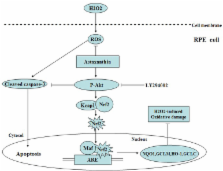
Oxidative stress on retinal pigment epithelial (RPE) cells is thought to play a crucial role in the development and progression of age-related macular degeneration. Astaxanthin (AST) is a carotenoid that shows significant antioxidant properties. This study was designed to investigate the protective effect of AST on ARPE-19 cells against oxidative stress and the possible underlying mechanism.
Methods
ARPE-19 cells exposed to different doses of H 2O 2 were incubated with various concentrations of AST and cell viability subsequently detected with the (4-[3-[4-iodophenyl]-2–4(4-nitrophenyl)-2H-5- tetrazolio-1,3-benzene disulfonate]; WST-1) assay. The apoptosis rate and intracellular levels of reactive oxygen species (ROS) were measured with flow cytometry. NAD(P)H quinine oxidoreductase 1 (NQO1), hemeoxygenase-1 (HO-1), glutamate-cysteine ligase modifier subunit (GCLM), and glutamate-cysteine ligase catalytic subunit (GCLC) expression were examined with real-time PCR and western blotting. The nuclear localization of nuclear factor (erythroid-derived 2)-like 2 (Nrf2) protein and the expression levels of cleaved caspase-3 and protein kinase B proteins were evaluated with western blotting.
Results
AST clearly reduced H 2O 2-induced cell viability loss, cell apoptosis, and intracellular generation of ROS. Furthermore, treatment with AST activated the Nrf2-ARE pathway by inducing Nrf2 nuclear localization. Consequently, Phase II enzymes NQO1, HO-1, GCLM, and GCLC mRNA and proteins were increased. AST inhibited expression of H 2O 2-induced cleaved caspase-3 protein. Activation of the phosphatidylinositol 3-kinase/protein kinase B (PI3K/Akt) pathway was involved in the protective effect of AST on the ARPE-19 cells.
Related collections
Most cited references30
- Record: found
- Abstract: found
- Article: not found
Astaxanthin: a review of its chemistry and applications.
- Record: found
- Abstract: found
- Article: not found
The role of oxidative stress in the pathogenesis of age-related macular degeneration.
- Record: found
- Abstract: not found
- Article: not found