8
views
0
recommends
+1 Recommend
0 collections
    0
    shares
      • Record: found
      • Abstract: found
      • Article: found
      Is Open Access

      Identification of a small molecule that primes the type I interferon response to cytosolic DNA

      research-article

      Read this article at

      Bookmark
          There is no author summary for this article yet. Authors can add summaries to their articles on ScienceOpen to make them more accessible to a non-specialist audience.

          Abstract

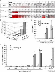
          The type I interferon response plays a pivotal role in host defense against infectious agents and tumors, and promising therapeutic approaches rely on small molecules designed to boost this system. To identify such compounds, we developed a high-throughput screening assay based on HEK-293 cells expressing luciferase under the control of Interferon-Stimulated Response Elements (ISRE). An original library of 10,000 synthetic compounds was screened, and we identified a series of 1H-benzimidazole-4-carboxamide compounds inducing the ISRE promoter sequence, specific cellular Interferon-Stimulated Genes (ISGs), and the phosphorylation of Interferon Regulatory Factor (IRF) 3. ISRE induction by ChX710, a prototypical member of this chemical series, was dependent on the adaptor MAVS and IRF1, but was IRF3 independent. Although it was unable to trigger type I IFN secretion per se, ChX710 efficiently primed cellular response to transfected plasmid DNA as assessed by potent synergistic effects on IFN-β secretion and ISG expression levels. This cellular response was dependent on STING, a key adaptor involved in the sensing of cytosolic DNA and immune activation by various pathogens, stress signals and tumorigenesis. Our results demonstrate that cellular response to cytosolic DNA can be boosted with a small molecule, and potential applications in antimicrobial and cancer therapies are discussed.

          Related collections

          Most cited references37

          • Record: found
          • Abstract: found
          • Article: not found

          Quantitative expression of toll-like receptor 1-10 mRNA in cellular subsets of human peripheral blood mononuclear cells and sensitivity to CpG oligodeoxynucleotides.

          The Toll-like receptor (TLR)9 is critical for the recognition of immunostimulatory CpG motifs but may cooperate with other TLRs. We analyzed TLR1-10 mRNA expression by using quantitative real-time PCR in highly purified subsets of human PBMC and determined the sensitivity of these subsets to CpG oligodeoxynucleotides (ODN). TLR1 and TLR6 were expressed in all cell types examined. TLR10 was highly expressed in B cells and weakly expressed in plasmacytoid dendritic cells (PDC). High expression of TLR2 was characteristic for monocytes. PDC and B cells expressed marked levels of TLR7 and TLR9 and were directly sensitive to CpG ODN. In CpG ODN-stimulated PDC and B cells, TLR9 expression rapidly decreased, as opposed to TLR7, which was up-regulated in PDC and decreased in B cells. In monocytes, NK cells, and T cells, TLR7 was absent. Despite low expression of TLR9, monocytes, NK cells, and T cells did not respond to CpG ODN in the absence of PDC but were activated in the presence of PDC. In conclusion, our studies provide evidence that PDC and B cells, but not monocytes, NK cells, or T cells, are primary targets of CpG ODN in peripheral blood. The characteristic expression pattern of TLR1-10 in cellular subsets of human PBMC is consistent with the concept that TLR9 is essential in the recognition of CpG ODN in PDC and B cells. In addition, selective regulation of TLR7 expression in PDC and B cells by CpG ODN revealed TLR7 as a candidate TLR potentially involved in modulating the recognition of CpG motifs.
            Bookmark
            • Record: found
            • Abstract: found
            • Article: not found

            DNA damage primes the type I interferon system via the cytosolic DNA sensor STING to promote anti-microbial innate immunity.

            Dysfunction in Ataxia-telangiectasia mutated (ATM), a central component of the DNA repair machinery, results in Ataxia Telangiectasia (AT), a cancer-prone disease with a variety of inflammatory manifestations. By analyzing AT patient samples and Atm(-/-) mice, we found that unrepaired DNA lesions induce type I interferons (IFNs), resulting in enhanced anti-viral and anti-bacterial responses in Atm(-/-) mice. Priming of the type I interferon system by DNA damage involved release of DNA into the cytoplasm where it activated the cytosolic DNA sensing STING-mediated pathway, which in turn enhanced responses to innate stimuli by activating the expression of Toll-like receptors, RIG-I-like receptors, cytoplasmic DNA sensors, and their downstream signaling partners. This study provides a potential explanation for the inflammatory phenotype of AT patients and establishes damaged DNA as a cell intrinsic danger signal that primes the innate immune system for a rapid and amplified response to microbial and environmental threats.
              Bookmark
              • Record: found
              • Abstract: found
              • Article: not found

              Peroxisomes are signaling platforms for antiviral innate immunity.

              Peroxisomes have long been established to play a central role in regulating various metabolic activities in mammalian cells. These organelles act in concert with mitochondria to control the metabolism of lipids and reactive oxygen species. However, while mitochondria have emerged as an important site of antiviral signal transduction, a role for peroxisomes in immune defense is unknown. Here, we report that the RIG-I-like receptor (RLR) adaptor protein MAVS is located on peroxisomes and mitochondria. We find that peroxisomal and mitochondrial MAVS act sequentially to create an antiviral cellular state. Upon viral infection, peroxisomal MAVS induces the rapid interferon-independent expression of defense factors that provide short-term protection, whereas mitochondrial MAVS activates an interferon-dependent signaling pathway with delayed kinetics, which amplifies and stabilizes the antiviral response. The interferon regulatory factor IRF1 plays a crucial role in regulating MAVS-dependent signaling from peroxisomes. These results establish that peroxisomes are an important site of antiviral signal transduction. Copyright (c) 2010 Elsevier Inc. All rights reserved.
                Bookmark

                Author and article information

                Contributors
                ftangy@pasteur.fr
                pierre-olivier.vidalain@parisdescartes.fr
                Journal
                Sci Rep
                Sci Rep
                Scientific Reports
                Nature Publishing Group UK (London )
                2045-2322
                31 May 2017
                31 May 2017
                2017
                : 7
                : 2561
                Affiliations
                [1 ]ISNI 0000 0001 2353 6535, GRID grid.428999.7, , Unité de Génomique Virale et Vaccination, Institut Pasteur, CNRS UMR3569, ; Paris, France
                [2 ]ISNI 0000 0001 2188 0914, GRID grid.10992.33, Mécanismes d’action des interférons et voies bio-thérapeutiques, , Université Paris Descartes, ; INSERM UMR-S1124, Paris, France
                [3 ]ISNI 0000 0001 2188 0914, GRID grid.10992.33, Chimie & Biologie, Modélisation et Immunologie pour la Thérapie (CBMIT), , Université Paris Descartes, CNRS UMR8601, ; Paris, France
                [4 ]ISNI 0000 0001 2353 6535, GRID grid.428999.7, Unité de Chimie et Biocatalyse, , Institut Pasteur, CNRS UMR3523, ; Paris, France
                [5 ]ISNI 0000 0001 2353 6535, GRID grid.428999.7, Unité de Virologie Moléculaire et Vaccinologie, , Institut Pasteur, ; Paris, France
                Author information
                http://orcid.org/0000-0001-9793-419X
                http://orcid.org/0000-0002-0202-612X
                Article
                2776
                10.1038/s41598-017-02776-z
                5451460
                28566766
                0ef7f68c-a6d1-4982-ab90-ddf381bd80ba
                © The Author(s) 2017

                Open Access This article is licensed under a Creative Commons Attribution 4.0 International License, which permits use, sharing, adaptation, distribution and reproduction in any medium or format, as long as you give appropriate credit to the original author(s) and the source, provide a link to the Creative Commons license, and indicate if changes were made. The images or other third party material in this article are included in the article’s Creative Commons license, unless indicated otherwise in a credit line to the material. If material is not included in the article’s Creative Commons license and your intended use is not permitted by statutory regulation or exceeds the permitted use, you will need to obtain permission directly from the copyright holder. To view a copy of this license, visit http://creativecommons.org/licenses/by/4.0/.

                History
                : 4 August 2016
                : 19 April 2017
                Categories
                Article
                Custom metadata
                © The Author(s) 2017

                Uncategorized
                Uncategorized

                Comments

                Comment on this article