8
views
0
recommends
+1 Recommend
0 collections
    0
    shares
      • Record: found
      • Abstract: found
      • Article: found
      Is Open Access

      Multispectral Fundus Photography of Choroidal Nevi with Trans-Palpebral Illumination

      Preprint
      research-article

      Read this article at

      Bookmark
          There is no author summary for this article yet. Authors can add summaries to their articles on ScienceOpen to make them more accessible to a non-specialist audience.

          Abstract

          Purpose:

          To investigate the spectral characteristics of choroidal nevi and assess the feasibility of quantifying the basal diameter of choroidal nevi using multispectral fundus images captured with trans-palpebral illumination.

          Methods:

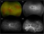
          The study employed a widefield fundus camera with multispectral (625 nm, 780 nm, 850 nm, and 970 nm) trans-palpebral illumination. Geometric features of choroidal nevi, including border clarity, overlying drusen, and lesion basal diameter, were characterized. Clinical imagers, including scanning laser ophthalmoscopy (SLO), autofluorescence (AF), and optical coherence tomography (OCT), were utilized for comparative assessment.

          Results:

          Fundus images captured with trans-palpebral illumination depicted nevi as dark regions with high contrast against the background. Near-infrared (NIR) fundus images provided enhanced visibility of lesion borders compared to visible light fundus images and SLO images. Lesion-background contrast measurements revealed 635 nm SLO at 11% and 625 nm fundus at 42%. Significantly enhanced contrasts were observed in NIR fundus images at 780 nm (73%), 850 nm (63%), and 970 nm (67%). For quantifying the basal diameter of nevi, NIR fundus images at 780 nm and 850 nm yielded a deviation of less than 10% when compared to OCT B-scan measurements.

          Conclusion:

          NIR fundus photography with trans-palpebral illumination enhances nevi visibility and boundary definition compared to SLO. Agreement in basal diameter measurements with OCT validates the accuracy and reliability of this method for choroidal nevi assessment.

          Translational Relevance:

          Multispectral fundus imaging with trans-palpebral illumination improves choroidal nevi visibility, accurately measures basal diameter, promising to enhance clinical practices in screening, diagnosis, and monitoring of choroidal nevi.

          Related collections

          Most cited references34

          • Record: found
          • Abstract: found
          • Article: found

          Fundus Photography in the 21st Century—A Review of Recent Technological Advances and Their Implications for Worldwide Healthcare

          The introduction of fundus photography has impacted retinal imaging and retinal screening programs significantly.
            Bookmark
            • Record: found
            • Abstract: found
            • Article: not found

            Therapeutic targets in age-related macular disease.

            Age-related macular disease (AMD) accounts for more than 50% of blind registration in Western society. Patients with AMD are classified as having early disease, in which visual function is well preserved, or late disease, in which central vision is lost. Until recently, there was no therapy available by which the course of the disorder could be modified. Now, the most common form of late-stage AMD - choroidal neovascularization - responds to treatment with anti-VEGF therapies; although visual loss is modified in a portion of these cases, no therapeutic approach exists that alters the evolution from early to late disease. However, as discussed in this Review, research over the last few years has demonstrated several features of AMD that are likely to be amenable to treatment. Potential targets for treatment are described, and possible therapeutic approaches are discussed.
              Bookmark
              • Record: found
              • Abstract: found
              • Article: found
              Is Open Access

              Ultra-widefield retinal imaging: an update on recent advances

              The development of ultra-widefield retinal imaging has accelerated our understanding of common retinal diseases. As we continue to validate the diagnostic and prognostic significance of pathology in the retinal periphery, the ability to visualize and evaluate these features in an efficient and patient-friendly manner will become more important. Current interest in ultra-widefield imaging includes the development of potential biomarkers of disease progression and indicators of preclinical disease development. This article reviews the current ultra-widefield imaging systems and recent advances in their applications to clinical practice with a focus on diabetic retinopathy, retinal vein occlusion, uveitis, and pediatric retina.
                Bookmark

                Author and article information

                Journal
                medRxiv
                MEDRXIV
                medRxiv
                Cold Spring Harbor Laboratory
                13 January 2024
                : 2024.01.12.24301119
                Affiliations
                [1 ]Department of Biomedical Engineering, University of Illinois Chicago, Chicago, IL 60607, USA
                [2 ]Department of Ophthalmology and Visual Sciences, University of Illinois Chicago, Chicago, IL, 60612, USA
                Author notes
                Corresponding author: Xincheng Yao, PhD, Richard & Loan Hill Chair Professor, Department of Biomedical Engineering (MC 563), University of Illinois Chicago, Clinical Sciences North, Suite W103, Room 164D, 820 South Wood Street, Chicago, IL 60612, Tel: (312)413-2016; Fax: (312)996-4644; xcy@ 123456uic.edu
                Article
                10.1101/2024.01.12.24301119
                10802649
                38260269
                0ec4a985-24c1-4e9f-b30d-4f3eed971824

                This work is licensed under a Creative Commons Attribution-NonCommercial-NoDerivatives 4.0 International License, which allows reusers to copy and distribute the material in any medium or format in unadapted form only, for noncommercial purposes only, and only so long as attribution is given to the creator.

                History
                Funding
                Funded by: National Eye Institute
                Award ID: R01 EY023522
                Award ID: R01 EY029673
                Award ID: R01 EY030101
                Award ID: R01 EY030842
                Award ID: P30EY001792
                Funded by: Research to Prevent Blindness
                Funded by: Richard and Loan Hill Endowment
                Categories
                Article

                Comments

                Comment on this article