25
views
0
recommends
+1 Recommend
0 collections
    0
    shares
      • Record: found
      • Abstract: found
      • Article: found
      Is Open Access

      Gene expression profiles in the bovine corpus luteum (CL) during the estrous cycle and pregnancy: Possible roles of chemokines in regulating CL function during pregnancy

      research-article

      Read this article at

      Bookmark
          There is no author summary for this article yet. Authors can add summaries to their articles on ScienceOpen to make them more accessible to a non-specialist audience.

          Abstract

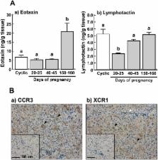
          To determine functional differences between the corpus luteum (CL) of the estrous cycle and pregnancy in cows, gene expression profiles were compared using a 15 K bovine oligo DNA microarray. In the pregnant CL at days 20–25, 40–45 and 150–160, the expressions of 138, 265 and 455 genes differed by a factor of > 2-fold (P < 0.05) from their expressions in the cyclic CL (days 10–12 of the estrous cycle). Messenger RNA expressions of chemokines ( eotaxin, lymphotactin and ENA-78) and their receptors ( CCR3, XCR1 and CXCR2) were validated by quantitative real-time PCR. Transcripts of eotaxin were more abundant in the CL at days 40–45 and 150–160 of pregnancy than in the cyclic CL (P < 0.01). In contrast, the mRNA expressions of lymphotactin, ENA-78 and XCR1 were lower in the CL of pregnancy (P < 0.05). Messenger RNAs of CCR3 and CXCR2 were similarly detected both in the cyclic and pregnant CL. Tissue protein levels of eotaxin were significantly higher in the CL at days 150–160 of pregnancy than in the CL at other stages, whereas the lymphotactin protein levels in the CL at days 20–25 of pregnancy were lower (P < 0.05). Immunohistochemical staining showed that CCR3 was expressed in the luteal cells and that XCR1 was expressed in both the luteal cells and endothelial cells. Collectively, the different gene expression profiles may contribute to functional differences between the cyclic and pregnant CL, and chemokines including eotaxin and lymphotactin may regulate CL function during pregnancy in cows.

          Related collections

          Most cited references38

          • Record: found
          • Abstract: found
          • Article: not found

          Lymphocyte traffic control by chemokines.

          In contrast to the remarkable chemokine responses of phagocytes and monocytes that were documented early on, lymphocytes have been considered for a long time to be poor targets for chemokine action. This view has changed dramatically with the discovery that peripheral blood T cells need to be activated before they can migrate in response to inflammatory chemokines. These chemokines do not act on the bulk of resting T cells that are in circulation. The identification of a new group of chemokines that selects resting, as opposed to effector, T and B cells was very exciting. These inflammation-unrelated chemokines affect transendothelial migration and localization of progenitor and mature lymphocytes in lymphoid and nonlymphoid tissues. Here, we summarize the current view of chemokine-mediated lymphocyte traffic and focus on the molecular mechanisms by which T cell responses to chemokines are modulated. Recent developments in this area justify the hypothesis that the distinct migration patterns of lymphocytes throughout their life cycle--that is, during lymphopoiesis, antigen-dependent priming, inflammation and immune surveillance--are finely tuned by changing sets of chemokines that are selective for developmentally regulated chemokine receptors. Thus, the chemokine system assures that cell traffic during inflammatory responses occurs in the proper spatial and temporal fashion and disturbance of this system, therefore, can lead to inflammatory disease.
            Bookmark
            • Record: found
            • Abstract: found
            • Article: not found

            Mechanisms controlling the function and life span of the corpus luteum.

            The primary function of the corpus luteum is secretion of the hormone progesterone, which is required for maintenance of normal pregnancy in mammals. The corpus luteum develops from residual follicular granulosal and thecal cells after ovulation. Luteinizing hormone (LH) from the anterior pituitary is important for normal development and function of the corpus luteum in most mammals, although growth hormone, prolactin, and estradiol also play a role in several species. The mature corpus luteum is composed of at least two steroidogenic cell types based on morphological and biochemical criteria and on the follicular source of origin. Small luteal cells appear to be of thecal cell origin and respond to LH with increased secretion of progesterone. LH directly stimulates the secretion of progesterone from small luteal cells via activation of the protein kinase A second messenger pathway. Large luteal cells are of granulosal cell origin and contain receptors for PGF(2alpha) and appear to mediate the luteolytic actions of this hormone. If pregnancy does not occur, the corpus luteum must regress to allow follicular growth and ovulation and the reproductive cycle begins again. Luteal regression is initiated by PGF(2alpha) of uterine origin in most subprimate species. The role played by PGF(2alpha) in primates remains controversial. In primates, if PGF(2alpha) plays a role in luteolysis, it appears to be of ovarian origin. The antisteroidogenic effects of PGF(2alpha) appear to be mediated by the protein kinase C second messenger pathway, whereas loss of luteal cells appears to follow an influx of calcium, activation of endonucleases, and an apoptotic form of cell death. If the female becomes pregnant, continued secretion of progesterone from the corpus luteum is required to provide an appropriate uterine environment for maintenance of pregnancy. The mechanisms whereby the pregnant uterus signals the corpus luteum that a conceptus is present varies from secretion of a chorionic gonadotropin (primates and equids), to secretion of an antiluteolytic factor (domestic ruminants), and to a neuroendocrine reflex arc that modifies the secretory patterns of hormones from the anterior pituitary (most rodents).
              Bookmark
              • Record: found
              • Abstract: found
              • Article: not found

              Expression of interferon (IFN)-stimulated genes in extrauterine tissues during early pregnancy in sheep is the consequence of endocrine IFN-tau release from the uterine vein.

              The ruminant conceptus synthesizes and secretes interferon (IFN)-tau, which presumably acts via an intrauterine paracrine mechanism to signal maternal recognition of pregnancy. The aims of this study were to determine whether IFN-stimulated genes (ISG) such as ISG15 and OAS-1 are differentially expressed in blood cells circulating in the uterus of ewes; whether extrauterine components of the reproductive tract such as the corpus luteum (CL) also express mRNA for these ISG, and whether antiviral activity is greater in uterine vein than in uterine artery during early pregnancy. The concentrations of mRNA for both ISG were significantly greater (P < 0.0001) in endometrium and jugular blood of 15-d pregnant ewes than in nonpregnant ewes. ISG15 and OAS-1 mRNA concentrations were also greater (P < 0.05) in CL from 15-d pregnant ewes than in nonpregnant ewes. Immunohistochemistry revealed intense staining for ISG15 in large luteal cells on d 15 of pregnancy. Blood cells from uterine artery and vein of 15-d pregnant ewes had similar ISG15 and OAS-1 mRNA concentrations, suggesting that these cells were not conditioned by IFN-tau within the uterus. By using an antiviral assay, uterine venous blood was found to contain 500- to 1000-fold higher concentrations of bioactive IFN-tau than in uterine arterial blood on d 15 of pregnancy. It is concluded that uterine vein releases IFN-tau, which induces ISG in extrauterine tissues such as the CL during the time of maternal recognition of pregnancy.
                Bookmark

                Author and article information

                Journal
                J Reprod Dev
                J. Reprod. Dev
                JRD
                The Journal of Reproduction and Development
                The Society for Reproduction and Development
                0916-8818
                1348-4400
                09 November 2014
                February 2015
                : 61
                : 1
                : 42-48
                Affiliations
                [1) ]Animal Physiology Research Unit, National Institute of Agrobiological Sciences, Ibaraki 305-0901, Japan
                [2) ]NARO Tohoku Agricultural Research Center, Iwate 020-0198, Japan
                [3) ]Laboratory of Veterinary Physiology, Iwate University, Iwate 020-8550, Japan
                [4) ]Laboratory of Reproductive Physiology, Okayama University, Okayama 700-8530, Japan
                Author notes
                Correspondence: R Sakumoto (e-mail: sakumoto@ 123456affrc.go.jp )
                Article
                2014-101
                10.1262/jrd.2014-101
                4354230
                25382605
                0dc81ec4-2d26-45d0-b36b-56211f79eb1b
                ©2015 Society for Reproduction and Development

                This is an open-access article distributed under the terms of the Creative Commons Attribution Non-Commercial No Derivatives (by-nc-nd) License.

                History
                : 23 August 2014
                : 14 October 2014
                Categories
                Original Article

                chemokines,corpus luteum,cow,microarray analysis,pregnancy
                chemokines, corpus luteum, cow, microarray analysis, pregnancy

                Comments

                Comment on this article