37
views
0
recommends
+1 Recommend
0 collections
    0
    shares
      • Record: found
      • Abstract: found
      • Article: found
      Is Open Access

      Cancer stem cell surface markers on normal stem cells

      review-article

      Read this article at

      Bookmark
          There is no author summary for this article yet. Authors can add summaries to their articles on ScienceOpen to make them more accessible to a non-specialist audience.

          Abstract

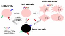
          The cancer stem cell (CSC) hypothesis has captured the attention of many scientists. It is believed that elimination of CSCs could possibly eradicate the whole cancer. CSC surface markers provide molecular targeted therapies for various cancers, using therapeutic antibodies specific for the CSC surface markers. Various CSC surface markers have been identified and published. Interestingly, most of the markers used to identify CSCs are derived from surface markers present on human embryonic stem cells (hESCs) or adult stem cells. In this review, we classify the currently known 40 CSC surface markers into 3 different categories, in terms of their expression in hESCs, adult stem cells, and normal tissue cells. Approximately 73% of current CSC surface markers appear to be present on embryonic or adult stem cells, and they are rarely expressed on normal tissue cells. The remaining CSC surface markers are considerably expressed even in normal tissue cells, and some of them have been extensively validated as CSC surface markers by various research groups. We discuss the significance of the categorized CSC surface markers, and provide insight into why surface markers on hESCs are an attractive source to find novel surface markers on CSCs.

          Related collections

          Most cited references118

          • Record: found
          • Abstract: found
          • Article: not found

          Identification of pancreatic cancer stem cells.

          Emerging evidence has suggested that the capability of a tumor to grow and propagate is dependent on a small subset of cells within a tumor, termed cancer stem cells. Although data have been provided to support this theory in human blood, brain, and breast cancers, the identity of pancreatic cancer stem cells has not been determined. Using a xenograft model in which primary human pancreatic adenocarcinomas were grown in immunocompromised mice, we identified a highly tumorigenic subpopulation of pancreatic cancer cells expressing the cell surface markers CD44, CD24, and epithelial-specific antigen (ESA). Pancreatic cancer cells with the CD44(+)CD24(+)ESA(+) phenotype (0.2-0.8% of pancreatic cancer cells) had a 100-fold increased tumorigenic potential compared with nontumorigenic cancer cells, with 50% of animals injected with as few as 100 CD44(+)CD24(+)ESA(+) cells forming tumors that were histologically indistinguishable from the human tumors from which they originated. The enhanced ability of CD44(+)CD24(+)ESA(+) pancreatic cancer cells to form tumors was confirmed in an orthotopic pancreatic tail injection model. The CD44(+)CD24(+)ESA(+) pancreatic cancer cells showed the stem cell properties of self-renewal, the ability to produce differentiated progeny, and increased expression of the developmental signaling molecule sonic hedgehog. Identification of pancreatic cancer stem cells and further elucidation of the signaling pathways that regulate their growth and survival may provide novel therapeutic approaches to treat pancreatic cancer, which is notoriously resistant to standard chemotherapy and radiation.
            Bookmark
            • Record: found
            • Abstract: found
            • Article: not found

            Identification of cells initiating human melanomas.

            Tumour-initiating cells capable of self-renewal and differentiation, which are responsible for tumour growth, have been identified in human haematological malignancies and solid cancers. If such minority populations are associated with tumour progression in human patients, specific targeting of tumour-initiating cells could be a strategy to eradicate cancers currently resistant to systemic therapy. Here we identify a subpopulation enriched for human malignant-melanoma-initiating cells (MMIC) defined by expression of the chemoresistance mediator ABCB5 (refs 7, 8) and show that specific targeting of this tumorigenic minority population inhibits tumour growth. ABCB5+ tumour cells detected in human melanoma patients show a primitive molecular phenotype and correlate with clinical melanoma progression. In serial human-to-mouse xenotransplantation experiments, ABCB5+ melanoma cells possess greater tumorigenic capacity than ABCB5- bulk populations and re-establish clinical tumour heterogeneity. In vivo genetic lineage tracking demonstrates a specific capacity of ABCB5+ subpopulations for self-renewal and differentiation, because ABCB5+ cancer cells generate both ABCB5+ and ABCB5- progeny, whereas ABCB5- tumour populations give rise, at lower rates, exclusively to ABCB5- cells. In an initial proof-of-principle analysis, designed to test the hypothesis that MMIC are also required for growth of established tumours, systemic administration of a monoclonal antibody directed at ABCB5, shown to be capable of inducing antibody-dependent cell-mediated cytotoxicity in ABCB5+ MMIC, exerted tumour-inhibitory effects. Identification of tumour-initiating cells with enhanced abundance in more advanced disease but susceptibility to specific targeting through a defining chemoresistance determinant has important implications for cancer therapy.
              Bookmark
              • Record: found
              • Abstract: found
              • Article: not found

              A Myc network accounts for similarities between embryonic stem and cancer cell transcription programs.

              c-Myc (Myc) is an important transcriptional regulator in embryonic stem (ES) cells, somatic cell reprogramming, and cancer. Here, we identify a Myc-centered regulatory network in ES cells by combining protein-protein and protein-DNA interaction studies and show that Myc interacts with the NuA4 complex, a regulator of ES cell identity. In combination with regulatory network information, we define three ES cell modules (Core, Polycomb, and Myc) and show that the modules are functionally separable, illustrating that the overall ES cell transcription program is composed of distinct units. With these modules as an analytical tool, we have reassessed the hypothesis linking an ES cell signature with cancer or cancer stem cells. We find that the Myc module, independent of the Core module, is active in various cancers and predicts cancer outcome. The apparent similarity of cancer and ES cell signatures reflects, in large part, the pervasive nature of Myc regulatory networks. Copyright © 2010 Elsevier Inc. All rights reserved.
                Bookmark

                Author and article information

                Journal
                BMB Rep
                BMB Rep
                BMB Reports
                Korean Society for Biochemistry and Molecular Biology
                1976-6696
                1976-670X
                June 2017
                30 June 2017
                : 50
                : 6
                : 285-298
                Affiliations
                Institute of Anticancer Medicine Development, Department of Integrative Bioscience and Biotechnology, Sejong University, Seoul 05006, Korea
                Author notes
                [* ]Corresponding author. Tel: +82-2-3408-3718; Fax: +82-2-3408- 4334; E-mail: cjryu@ 123456sejong.ac.kr
                Article
                bmb-50-285
                10.5483/BMBRep.2017.50.6.039
                5498139
                28270302
                0cc6f17e-3984-4bf1-9220-e91c1ee719d6
                Copyright © 2017 by the The Korean Society for Biochemistry and Molecular Biology

                This is an open-access article distributed under the terms of the Creative Commons Attribution Non-Commercial License ( http://creativecommons.org/licenses/by-nc/4.0) which permits unrestricted non-commercial use, distribution, and reproduction in any medium, provided the original work is properly cited.

                History
                : 07 March 2017
                Categories
                Invited Mini Review

                adult stem cells,cancer stem cells,human embryonic stem cells,normal tissue cells,surface marker

                Comments

                Comment on this article