13
views
0
recommends
+1 Recommend
0 collections
    0
    shares
      • Record: found
      • Abstract: found
      • Article: found
      Is Open Access

      Tor1a+/- mice develop dystonia-like movements via a striatal dopaminergic dysregulation triggered by peripheral nerve injury

      research-article

      Read this article at

      Bookmark
          There is no author summary for this article yet. Authors can add summaries to their articles on ScienceOpen to make them more accessible to a non-specialist audience.

          Abstract

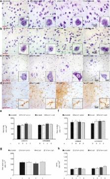
          Isolated generalized dystonia is a central motor network disorder characterized by twisted movements or postures. The most frequent genetic cause is a GAG deletion in the Tor1a (DYT1) gene encoding torsinA with a reduced penetrance of 30-40 % suggesting additional genetic or environmental modifiers. Development of dystonia-like movements after a standardized peripheral nerve crush lesion in wild type (wt) and Tor1a+/- mice, that express 50 % torsinA only, was assessed by scoring of hindlimb movements during tail suspension, by rotarod testing and by computer-assisted gait analysis. Western blot analysis was performed for dopamine transporter (DAT), D1 and D2 receptors from striatal and quantitative RT-PCR analysis for DAT from midbrain dissections. Autoradiography was used to assess the functional DAT binding in striatum. Striatal dopamine and its metabolites were analyzed by high performance liquid chromatography. After nerve crush injury, we found abnormal posturing in the lesioned hindlimb of both mutant and wt mice indicating the profound influence of the nerve lesion (15x vs. 12x relative to control) resembling human peripheral pseudodystonia. In mutant mice the phenotypic abnormalities were increased by about 40 % ( p < 0.05). This was accompanied by complex alterations of striatal dopamine homeostasis. Pharmacological blockade of dopamine synthesis reduced severity of dystonia-like movements, whereas treatment with L-Dopa aggravated these but only in mutant mice suggesting a DYT1 related central component relevant to the development of abnormal involuntary movements. Our findings suggest that upon peripheral nerve injury reduced torsinA concentration and environmental stressors may act in concert in causing the central motor network dysfunction of DYT1 dystonia.

          Electronic supplementary material

          The online version of this article (doi:10.1186/s40478-016-0375-7) contains supplementary material, which is available to authorized users.

          Related collections

          Most cited references53

          • Record: found
          • Abstract: found
          • Article: not found

          The functional neuroanatomy of dystonia.

          Dystonia is a neurological disorder characterized by involuntary twisting movements and postures. There are many different clinical manifestations, and many different causes. The neuroanatomical substrates for dystonia are only partly understood. Although the traditional view localizes dystonia to basal ganglia circuits, there is increasing recognition that this view is inadequate for accommodating a substantial portion of available clinical and experimental evidence. A model in which several brain regions play a role in a network better accommodates the evidence. This network model accommodates neuropathological and neuroimaging evidence that dystonia may be associated with abnormalities in multiple different brain regions. It also accommodates animal studies showing that dystonic movements arise with manipulations of different brain regions. It is consistent with neurophysiological evidence suggesting defects in neural inhibitory processes, sensorimotor integration, and maladaptive plasticity. Finally, it may explain neurosurgical experience showing that targeting the basal ganglia is effective only for certain subpopulations of dystonia. Most importantly, the network model provides many new and testable hypotheses with direct relevance for new treatment strategies that go beyond the basal ganglia. This article is part of a Special Issue entitled "Advances in dystonia". Copyright © 2011 Elsevier Inc. All rights reserved.
            Bookmark
            • Record: found
            • Abstract: found
            • Article: not found

            Loss of the dystonia-associated protein torsinA selectively disrupts the neuronal nuclear envelope.

            An enigmatic feature of many genetic diseases is that mutations in widely expressed genes cause tissue-specific illness. One example is DYT1 dystonia, a neurodevelopmental disease caused by an in-frame deletion (Deltagag) in the gene encoding torsinA. Here we show that neurons from both torsinA null (Tor1a(-/-)) and homozygous disease mutant "knockin" mice (Tor1a(Deltagag/Deltagag)) contain severely abnormal nuclear membranes, although non-neuronal cell types appear normal. These membrane abnormalities develop in postmigratory embryonic neurons and subsequently worsen with further neuronal maturation, a finding evocative of the developmental dependence of DYT1 dystonia. These observations demonstrate that neurons have a unique requirement for nuclear envelope localized torsinA function and suggest that loss of this activity is a key molecular event in the pathogenesis of DYT1 dystonia.
              Bookmark
              • Record: found
              • Abstract: found
              • Article: not found

              The neural substrates of Rapid-Onset Dystonia-Parkinsonism

              Although dystonias are a common group of movement disorders the mechanisms by which brain dysfunction results in dystonia are not understood. Rapid-onset Dystonia-Parkinsonism is a hereditary dystonia caused by mutations in the ATP1A3 gene. Affected subjects can be symptom free for years but rapidly develop persistent dystonia and parkinsonism-like symptoms after a stressful experience. Using a mouse model here we show that an adverse interaction between the cerebellum and basal ganglia can account for the symptoms of the patients. The primary instigator of dystonia is the cerebellum whose aberrant activity alters basal ganglia function which in turn causes dystonia. This adverse interaction between the cerebellum and basal ganglia is mediated through a di-synaptic thalamic pathway which when severed is effective in alleviating dystonia. Our results provide a unifying hypothesis for the involvement of cerebellum and basal ganglia in generation of dystonia and suggest therapeutic strategies for the treatment of RDP.
                Bookmark

                Author and article information

                Contributors
                +49 931 20124719 , ip_c@ukw.de
                Journal
                Acta Neuropathol Commun
                Acta Neuropathol Commun
                Acta Neuropathologica Communications
                BioMed Central (London )
                2051-5960
                3 October 2016
                3 October 2016
                2016
                : 4
                : 108
                Affiliations
                [1 ]Department of Neurology, University Hospital of Würzburg, University of Würzburg, Josef-Schneider-Strasse 11, 97080 Würzburg, Germany
                [2 ]Department of Psychiatry, Psychosomatics and Psychotherapy, Goethe-University Frankfurt, Frankfurt am Main, Germany
                [3 ]Department of Nuclear Medicine, University Hospital of Würzburg, University of Würzburg, Oberdürrbacher Strasse 6, 97080 Würzburg, Germany
                [4 ]Toronto Western Hospital, University Health Network, 100 King St W, Suite 5600, Toronto, Canada
                Author information
                http://orcid.org/0000-0003-0484-385X
                Article
                375
                10.1186/s40478-016-0375-7
                5048687
                27716431
                0bc904de-f36c-403f-a958-a75e841ccbb3
                © The Author(s). 2016

                Open AccessThis article is distributed under the terms of the Creative Commons Attribution 4.0 International License ( http://creativecommons.org/licenses/by/4.0/), which permits unrestricted use, distribution, and reproduction in any medium, provided you give appropriate credit to the original author(s) and the source, provide a link to the Creative Commons license, and indicate if changes were made. The Creative Commons Public Domain Dedication waiver ( http://creativecommons.org/publicdomain/zero/1.0/) applies to the data made available in this article, unless otherwise stated.

                History
                : 8 September 2016
                : 14 September 2016
                Funding
                Funded by: FundRef http://dx.doi.org/10.13039/501100002347, Bundesministerium für Bildung und Forschung;
                Award ID: DYSTRACT
                Award Recipient :
                Funded by: FundRef http://dx.doi.org/10.13039/501100001659, Deutsche Forschungsgemeinschaft;
                Award ID: TRR SFB 58, B06
                Award Recipient :
                Funded by: State of Bavaria
                Categories
                Research
                Custom metadata
                © The Author(s) 2016

                dystonia,dyt1,dopamine,peripheral injury,second hit
                dystonia, dyt1, dopamine, peripheral injury, second hit

                Comments

                Comment on this article