2
views
0
recommends
+1 Recommend
0 collections
    0
    shares
      • Record: found
      • Abstract: found
      • Article: found
      Is Open Access

      Calcified Leiomyoma of the Distal Forearm in a Child: A Case Report and Review of Literature

      case-report

      Read this article at

      Bookmark
          There is no author summary for this article yet. Authors can add summaries to their articles on ScienceOpen to make them more accessible to a non-specialist audience.

          Abstract

          Background

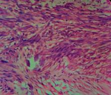
          Deep somatic leiomyomas arising in skeletal muscle are extremely rare in children, especially in the extremities. Around half of them show calcifications. We present a rare case of a calcified leiomyoma of the distal forearm in a child. Case Summary. A seven-year-old boy presented with right distal forearm and wrist pain with restricted supination for 4 years. X-ray showed ring and arc calcifications in the distal forearm at the interosseous area. MRI also confirmed a well-defined soft tissue lesion with areas of calcifications. A diagnosis of a cartilage-forming lesion or a peripheral nerve sheath tumour was suggested. The lesion was completely excised. Histology showed a lesion composed of intersecting fascicles of spindle cells with stromal calcification having immunohistochemical features of a leiomyoma.

          Conclusion

          Although soft tissue calcifications can be seen in a plethora of conditions seen in daily orthopaedic practise, a high index of suspicion should be maintained for rare conditions like deep somatic leiomyoma.

          Related collections

          Most cited references16

          • Record: found
          • Abstract: found
          • Article: not found

          CARE 2013 Explanations and Elaborations: Reporting Guidelines for Case Reports.

          Well-written and transparent case reports (1) reveal early signals of potential benefits, harms, and information on the use of resources; (2) provide information for clinical research and clinical practice guidelines (CPGs), and (3) inform medical education. High-quality case reports are more likely when authors follow reporting guidelines. During 2011-2012 a group of clinicians, researchers, and journal editors developed recommendations for the accurate reporting of information in case reports that resulted in the CARE (CAse REport) Statement and Checklist. They were presented at the 2013 International Congress on Peer Review and Biomedical Publication, have been endorsed by multiple medical journals, and translated into nine languages. This explanation and elaboration document has the objective to increase the use and dissemination of the CARE Checklist in writing and publishing case reports. Each item from the CARE Checklist is explained and accompanied by published examples. The explanations and examples in this document are designed to support the writing of high-quality case reports by authors and their critical appraisal by editors, peer reviewers, and readers. This article and the 2013 CARE Statement and Checklist, available from the CARE website [www.care-statement.org] and the EQUATOR Network, [www.equator-network.org] are resources for improving the completeness and transparency of case reports.
            Bookmark
            • Record: found
            • Abstract: found
            • Article: not found

            Smooth muscle tumors of soft tissue.

            Jie Weiss (2002)
            This paper presents an overview of smooth muscle tumors occurring in deep soft tissue. Although the existence of leiomyomas of soft tissue has been questioned in the past, it appears that they do exist but are rare, and must be diagnosed using stringent histologic criteria that include no atypia and minimal or no mitotic activity. They segregate into two distinct clinicopathologic groups, one group occurring in patients of either sex in deep somatic soft tissue and the second occurring primarily in women in the pelvic retroperitoneum. The latter bear a histologic similarity to uterine leiomyomas. Leiomyosarcomas occur in retroperitoneum followed by deep somatic soft tissue and are diagnosed by the presence of nuclear atypia and essentially any level of mitotic activity. Leiomyosarcomas of deep somatic tissue commonly arise from small veins and their behavior can be predicted by a number of factors including age, grade, and "disruption" of tumor. Conversely, few factors have proved to be prognostically useful for leiomyosarcomas of the retroperitoneum, as nearly all prove fatal. Epstein Barr virus (EBV)-associated smooth muscle tumors are a recently emerging entity that occur in the setting of immunocompromise. Their behavior is closely tied to the immune status of the patient rather than to specific histologic features.
              Bookmark
              • Record: found
              • Abstract: found
              • Article: not found

              Malignant peripheral nerve sheath tumors (malignant schwannomas) in children.

              We studied the clinical and pathologic features of 78 malignant peripheral nerve sheath tumors in children less than or equal to 15 years of age. There were 42 boys and 36 girls, with a median age of 10 years. The majority of the tumors (42, or 54%) were central or axial in location; the rest were peripheral. Sixteen patients (21%) had a history of von Recklinghausen's disease. Fourteen (18%) had a malignant peripheral nerve sheath tumor arising in a nerve trunk or a neurofibroma and were unassociated with von Recklinghausen's disease. Patients typically presented with a painful mass of variable duration. Tumors ranged from 2 to 33 cm (median, 7.5 cm) and demonstrated a wide histologic spectrum that included spindled, epithelioid, and primitive neuroepithelial-like cells as well as heterologous elements (11). Immunohistochemical staining revealed S-100 protein in 28 of 50 cases (56%) as well as vimentin (13 of 21 cases, or 62%), Leu 7 (22 of 49 cases, or 45%), actin (eight of 20 cases, or 40%), and keratin (seven of 27 cases, or 26%). Survival status was known for 57 patients (73%). Kaplan-Meier estimates revealed a median survival of 45 months. Half of the patients had local recurrences at 12 months, and half had metastases at 24 months, most commonly to lungs, followed by lymph nodes, liver, bone, soft tissue, and brain. Age greater than or equal to 7 years, male sex, presence of von Recklinghausen's disease, central location, larger tumor size, and tumors with greater than or equal to 25% necrosis were found to be potentially significant adverse prognostic indicators by univariate analysis. Multivariate analysis revealed that larger tumor size, age greater than or equal to 7 years, tumor necrosis greater than or equal to 25%, and von Recklinghausen's disease to be independent adverse prognostic factors.
                Bookmark

                Author and article information

                Contributors
                Journal
                Case Rep Orthop
                Case Rep Orthop
                CRIOR
                Case Reports in Orthopedics
                Hindawi
                2090-6749
                2090-6757
                2020
                28 August 2020
                : 2020
                : 8821265
                Affiliations
                1National Hospital of Sri Lanka, Colombo, Sri Lanka
                2Lady Ridgeway Hospital for Children, Colombo, Sri Lanka
                3Colombo North Teaching Hospital, Ragama, Sri Lanka
                Author notes

                Academic Editor: Koichi Sairyo

                Author information
                https://orcid.org/0000-0002-2010-8460
                Article
                10.1155/2020/8821265
                7475744
                32908750
                0bbf1837-b241-4d0c-bea8-21435c85b704
                Copyright © 2020 Nadeeke Nidhan Perera et al.

                This is an open access article distributed under the Creative Commons Attribution License, which permits unrestricted use, distribution, and reproduction in any medium, provided the original work is properly cited.

                History
                : 10 May 2020
                : 17 August 2020
                : 24 August 2020
                Categories
                Case Report

                Orthopedics
                Orthopedics

                Comments

                Comment on this article