11
views
0
recommends
+1 Recommend
0 collections
    0
    shares
      • Record: found
      • Abstract: found
      • Article: found
      Is Open Access

      Complete human day 14 post-implantation embryo models from naive ES cells

      research-article

      Read this article at

      Bookmark
          There is no author summary for this article yet. Authors can add summaries to their articles on ScienceOpen to make them more accessible to a non-specialist audience.

          Abstract

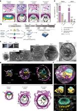
          The ability to study human post-implantation development remains limited owing to ethical and technical challenges associated with intrauterine development after implantation 1 . Embryo-like models with spatially organized morphogenesis and structure of all defining embryonic and extra-embryonic tissues of the post-implantation human conceptus (that is, the embryonic disc, the bilaminar disc, the yolk sac, the chorionic sac and the surrounding trophoblast layer) remain lacking 1, 2 . Mouse naive embryonic stem cells have recently been shown to give rise to embryonic and extra-embryonic stem cells capable of self-assembling into post-gastrulation structured stem-cell-based embryo models with spatially organized morphogenesis (called SEMs) 3 . Here we extend those findings to humans using only genetically unmodified human naive embryonic stem cells (cultured in human enhanced naive stem cell medium conditions) 4 . Such human fully integrated and complete SEMs recapitulate the organization of nearly all known lineages and compartments of post-implantation human embryos, including the epiblast, the hypoblast, the extra-embryonic mesoderm and the trophoblast layer surrounding the latter compartments. These human complete SEMs demonstrated developmental growth dynamics that resemble key hallmarks of post-implantation stage embryogenesis up to 13–14 days after fertilization (Carnegie stage 6a). These include embryonic disc and bilaminar disc formation, epiblast lumenogenesis, polarized amniogenesis, anterior–posterior symmetry breaking, primordial germ-cell specification, polarized yolk sac with visceral and parietal endoderm formation, extra-embryonic mesoderm expansion that defines a chorionic cavity and a connecting stalk, and a trophoblast-surrounding compartment demonstrating syncytium and lacunae formation. This SEM platform will probably enable the experimental investigation of previously inaccessible windows of human early post implantation up to peri-gastrulation development.

          Abstract

          The culture of genetically unmodified human naive embryonic stem cells in specific growth conditions gives rise to structures that recapitulate those of post-implantation human embryos up to 13–14 days after fertilization.

          Related collections

          Most cited references44

          • Record: found
          • Abstract: found
          • Article: not found

          Comprehensive Integration of Single-Cell Data

          Single-cell transcriptomics has transformed our ability to characterize cell states, but deep biological understanding requires more than a taxonomic listing of clusters. As new methods arise to measure distinct cellular modalities, a key analytical challenge is to integrate these datasets to better understand cellular identity and function. Here, we develop a strategy to "anchor" diverse datasets together, enabling us to integrate single-cell measurements not only across scRNA-seq technologies, but also across different modalities. After demonstrating improvement over existing methods for integrating scRNA-seq data, we anchor scRNA-seq experiments with scATAC-seq to explore chromatin differences in closely related interneuron subsets and project protein expression measurements onto a bone marrow atlas to characterize lymphocyte populations. Lastly, we harmonize in situ gene expression and scRNA-seq datasets, allowing transcriptome-wide imputation of spatial gene expression patterns. Our work presents a strategy for the assembly of harmonized references and transfer of information across datasets.
            Bookmark
            • Record: found
            • Abstract: found
            • Article: not found

            Integrating single-cell transcriptomic data across different conditions, technologies, and species

            Computational single-cell RNA-seq (scRNA-seq) methods have been successfully applied to experiments representing a single condition, technology, or species to discover and define cellular phenotypes. However, identifying subpopulations of cells that are present across multiple data sets remains challenging. Here, we introduce an analytical strategy for integrating scRNA-seq data sets based on common sources of variation, enabling the identification of shared populations across data sets and downstream comparative analysis. We apply this approach, implemented in our R toolkit Seurat (http://satijalab.org/seurat/), to align scRNA-seq data sets of peripheral blood mononuclear cells under resting and stimulated conditions, hematopoietic progenitors sequenced using two profiling technologies, and pancreatic cell 'atlases' generated from human and mouse islets. In each case, we learn distinct or transitional cell states jointly across data sets, while boosting statistical power through integrated analysis. Our approach facilitates general comparisons of scRNA-seq data sets, potentially deepening our understanding of how distinct cell states respond to perturbation, disease, and evolution.
              Bookmark
              • Record: found
              • Abstract: found
              • Article: not found

              Batch effects in single-cell RNA-sequencing data are corrected by matching mutual nearest neighbors

              Large-scale single-cell RNA sequencing (scRNA-seq) datasets that are produced in different laboratories and at different times contain batch effects that could compromise integration and interpretation of these data. Existing scRNA-seq analysis methods incorrectly assume that the composition of cell populations is either known, or the same, across batches. We present a strategy for batch correction that is based on the detection of mutual nearest neighbours (MNN) in the high-dimensional expression space. Our approach does not rely on pre-defined or equal population compositions across batches, and only requires that a subset of the population be shared between batches. We demonstrate the superiority of our approach over existing methods using both simulated and real scRNA-seq data sets. Using multiple droplet-based scRNA-seq data sets, we demonstrate that our MNN batch-effect correction method scales to large numbers of cells.
                Bookmark

                Author and article information

                Contributors
                jacob.hanna@weizmann.ac.il
                Journal
                Nature
                Nature
                Nature
                Nature Publishing Group UK (London )
                0028-0836
                1476-4687
                6 September 2023
                6 September 2023
                2023
                : 622
                : 7983
                : 562-573
                Affiliations
                [1 ]Department of Molecular Genetics, Weizmann Institute of Science, ( https://ror.org/0316ej306) Rehovot, Israel
                [2 ]Department of Clinical Sciences, Intervention and Technology, Karolinska Institutet, ( https://ror.org/056d84691) Stockholm, Sweden
                [3 ]Division of Obstetrics and Gynecology, Karolinska Universitetssjukhuset, ( https://ror.org/00m8d6786) Stockholm, Sweden
                [4 ]Department of Development and Regeneration, Leuven Stem Cell Institute, Leuven Institute for Single-cell Omics (LISCO), KU Leuven-University of Leuven, ( https://ror.org/05f950310) Leuven, Belgium
                [5 ]Department of Life Sciences Core Facilities, Weizmann Institute of Science, ( https://ror.org/0316ej306) Rehovot, Israel
                [6 ]Département de Médecine, Université de Montreal, ( https://ror.org/0161xgx34) Montreal, Quebec Canada
                [7 ]GRID grid.14848.31, ISNI 0000 0001 2292 3357, Centre de Recherche du Centre, , Hospitalier de l’Université de Montréal Axe Immunopathologie, ; Montreal, Quebec Canada
                [8 ]Ming Wai Lau Center for Reparative Medicine, Stockholm Node, Karolinska Institutet, ( https://ror.org/056d84691) Stockholm, Sweden
                Author information
                http://orcid.org/0000-0002-1339-7778
                http://orcid.org/0000-0002-4121-4289
                http://orcid.org/0000-0002-4878-2174
                http://orcid.org/0000-0001-9827-0436
                http://orcid.org/0000-0002-2771-7445
                http://orcid.org/0000-0002-2244-6877
                http://orcid.org/0000-0003-2042-9974
                Article
                6604
                10.1038/s41586-023-06604-5
                10584686
                37673118
                0af87de5-dcff-430d-b347-d98161d3625d
                © The Author(s) 2023

                Open Access This article is licensed under a Creative Commons Attribution 4.0 International License, which permits use, sharing, adaptation, distribution and reproduction in any medium or format, as long as you give appropriate credit to the original author(s) and the source, provide a link to the Creative Commons licence, and indicate if changes were made. The images or other third party material in this article are included in the article’s Creative Commons licence, unless indicated otherwise in a credit line to the material. If material is not included in the article’s Creative Commons licence and your intended use is not permitted by statutory regulation or exceeds the permitted use, you will need to obtain permission directly from the copyright holder. To view a copy of this licence, visit http://creativecommons.org/licenses/by/4.0/.

                History
                : 11 April 2023
                : 4 September 2023
                Categories
                Article
                Custom metadata
                © Springer Nature Limited 2023

                Uncategorized
                induced pluripotent stem cells,reproductive biology,differentiation,embryonic induction,embryology

                Comments

                Comment on this article