36
views
0
recommends
+1 Recommend
0 collections
    0
    shares
      • Record: found
      • Abstract: found
      • Article: found
      Is Open Access

      Mitochondria in endothelial cells: Sensors and integrators of environmental cues

      review-article
      a , a , b , *
      Redox Biology
      Elsevier
      Endothelial cells, Mitochondria, Biogenesis, Dynamics, ROS

      Read this article at
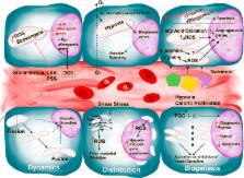
      Bookmark
          There is no author summary for this article yet. Authors can add summaries to their articles on ScienceOpen to make them more accessible to a non-specialist audience.

          Abstract

          The involvement of angiogenesis in disease and its potential as a therapeutic target have been firmly established over recent decades. Endothelial cells (ECs) are central elements in vessel homeostasis and regulate the passage of material and cells into and out of the bloodstream. EC proliferation and migration are modified by alterations to mitochondrial biogenesis and dynamics resulting from several signals and environmental cues, such as oxygen, hemodynamics, and nutrients. As intermediary signals, mitochondrial ROS are released as important downstream modulators of the expression of angiogenesis-related genes. In this review, we discuss the physiological actions of these signals and aberrant responses during vascular disorders.

          Highlights

          • Mitochondria in EC act as integrators of environmental cues.

          • Circulating signals modify mitochondrial dynamics, altering EC phenotype.

          • ROS release by EC mitochondria regulates expression of vascular genes.

          Related collections

          Most cited references90

          • Record: found
          • Abstract: found
          • Article: not found

          Mitochondrial dynamics and apoptosis.

          In healthy cells, mitochondria continually divide and fuse to form a dynamic interconnecting network. The molecular machinery that mediates this organelle fission and fusion is necessary to maintain mitochondrial integrity, perhaps by facilitating DNA or protein quality control. This network disintegrates during apoptosis at the time of cytochrome c release and prior to caspase activation, yielding more numerous and smaller mitochondria. Recent work shows that proteins involved in mitochondrial fission and fusion also actively participate in apoptosis induction. This review will cover the recent advances and presents competing models on how the mitochondrial fission and fusion machinery may intersect apoptosis pathways.
            Bookmark
            • Record: found
            • Abstract: found
            • Article: not found

            Links between metabolism and cancer.

            Chi Dang (2012)
            Metabolism generates oxygen radicals, which contribute to oncogenic mutations. Activated oncogenes and loss of tumor suppressors in turn alter metabolism and induce aerobic glycolysis. Aerobic glycolysis or the Warburg effect links the high rate of glucose fermentation to cancer. Together with glutamine, glucose via glycolysis provides the carbon skeletons, NADPH, and ATP to build new cancer cells, which persist in hypoxia that in turn rewires metabolic pathways for cell growth and survival. Excessive caloric intake is associated with an increased risk for cancers, while caloric restriction is protective, perhaps through clearance of mitochondria or mitophagy, thereby reducing oxidative stress. Hence, the links between metabolism and cancer are multifaceted, spanning from the low incidence of cancer in large mammals with low specific metabolic rates to altered cancer cell metabolism resulting from mutated enzymes or cancer genes.
              Bookmark
              • Record: found
              • Abstract: found
              • Article: not found

              Fatty acid carbon is essential for dNTP synthesis in endothelial cells

              The metabolism of endothelial cells (ECs) during vessel sprouting remains poorly studied. Here, we report that endothelial loss of CPT1a, a rate-limiting enzyme of fatty acid oxidation (FAO), caused vascular sprouting defects due to impaired proliferation, not migration of ECs. Reduction of FAO in ECs did not cause energy depletion or disturb redox homeostasis, but impaired de novo nucleotide synthesis for DNA replication. Isotope labeling studies in control ECs showed that fatty acid carbons substantially replenished the Krebs cycle, and were incorporated into aspartate (a nucleotide precursor), uridine monophosphate (a precursor of pyrimidine nucleoside triphosphates) and DNA. CPT1a silencing reduced these processes and depleted EC stores of aspartate and deoxyribonucleoside triphosphates. Acetate (metabolized to acetyl-CoA, thereby substituting for the depleted FAO-derived acetyl-CoA) or a nucleoside mix rescued the phenotype of CPT1a-silenced ECs. Finally, CPT1 blockade inhibited pathological ocular angiogenesis, suggesting a novel strategy for blocking angiogenesis.
                Bookmark

                Author and article information

                Contributors
                Journal
                Redox Biol
                Redox Biol
                Redox Biology
                Elsevier
                2213-2317
                18 April 2017
                August 2017
                18 April 2017
                : 12
                : 821-827
                Affiliations
                [a ]Centro Nacional de Investigaciones Cardiovasculares Carlos III (CNIC), Melchor Fernández Almagro 3, 28029 Madrid, Spain
                [b ]Centro de Investigaciones en RED (CIBERFES), Melchor Fernández Almagro, 28029 Madrid, Spain
                Author notes
                [* ]Corresponding author at: Centro Nacional de Investigaciones Cardiovasculares Carlos III (CNIC), Melchor Fernández Almagro 3, 28029 Madrid, Spain. jaenriquez@ 123456cnic.es
                Article
                S2213-2317(17)30110-6
                10.1016/j.redox.2017.04.021
                5406579
                28448943
                0ac09268-8c0a-4dc1-830a-ab64e61e0b4a
                © 2017 The Authors

                This is an open access article under the CC BY-NC-ND license (http://creativecommons.org/licenses/by-nc-nd/4.0/).

                History
                : 11 February 2017
                : 23 March 2017
                : 13 April 2017
                Categories
                Review Article

                endothelial cells,mitochondria,biogenesis,dynamics,ros
                endothelial cells, mitochondria, biogenesis, dynamics, ros

                Comments

                Comment on this article