0
views
0
recommends
+1 Recommend
0 collections
    0
    shares
      • Record: found
      • Abstract: found
      • Article: found
      Is Open Access

      HPV status and immunohistochemical analysis of p16, p53 and PD‑L1 expression as prognostic biomarkers in patients with squamous cell anal cancer receiving definitive radiotherapy/chemoradiotherapy

      research-article

      Read this article at

      Bookmark
          There is no author summary for this article yet. Authors can add summaries to their articles on ScienceOpen to make them more accessible to a non-specialist audience.

          Abstract

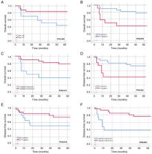
          Anal squamous cell carcinoma (SCC) treated with definitive radiotherapy (RT)/chemoradiotherapy (CRT) has shown high success rates, yet challenges such as treatment resistance and recurrence persist. The present study aimed to investigate the associations between immunohistochemical (IHC) evaluation, treatment response and prognosis in anal SCC. A retrospective cohort analysis included 42 patients with anal SCC treated at a single institution between 2006 and 2022. Human papillomavirus (HPV) status was determined, and the IHC analysis of p16, p53 and PD-L1 expression was conducted using formalin-fixed, paraffin-embedded biopsies. A complete response to RT/CRT was observed in 71.4% of patients. Recurrence occurred in 38.1% of cases, of which 7.1% had local-regional recurrence (LRR), 14.3% had distant recurrence (DR), and 16.7% had both LRR and DR. HPV positivity (71.4%) was significantly associated with p16 positivity. Lack of complete response was associated with HPV-negative status, p16-negative status, increased recurrence and DR. In addition, recurrence was significantly associated with p53-positive status, and p53 positivity was significantly associated with increased LRR. PD-L1 positivity, defined as a combined positive score (CPS) ≥1% was found in 73.8% of the patients, and exhibited significant associations with HPV positivity and p16 positivity. PD-L1 CPS ≥ 1% was also associated with an increased LRR. Univariate analysis revealed that age <65 years, a complete response and HPV positivity were associated with increased 5-year overall survival (OS), while a complete response, HPV positivity and p53-negative status were associated with increased 5-year disease-free survival (DFS). Multivariate analysis identified that age <65 years and HPV positivity are independent prognostic factors for 5-year OS, and a complete response and p53-negative status are independent prognostic factors for 5-year DFS. In conclusion, these findings suggust that the identification of HPV status and poor prognostic biomarkers at diagnosis may be used to guide personalized treatment strategies, with the combination of immunotherapy with standard CRT potentially providing improved outcomes.

          Related collections

          Most cited references64

          • Record: found
          • Abstract: found
          • Article: not found

          New response evaluation criteria in solid tumours: revised RECIST guideline (version 1.1).

          Assessment of the change in tumour burden is an important feature of the clinical evaluation of cancer therapeutics: both tumour shrinkage (objective response) and disease progression are useful endpoints in clinical trials. Since RECIST was published in 2000, many investigators, cooperative groups, industry and government authorities have adopted these criteria in the assessment of treatment outcomes. However, a number of questions and issues have arisen which have led to the development of a revised RECIST guideline (version 1.1). Evidence for changes, summarised in separate papers in this special issue, has come from assessment of a large data warehouse (>6500 patients), simulation studies and literature reviews. HIGHLIGHTS OF REVISED RECIST 1.1: Major changes include: Number of lesions to be assessed: based on evidence from numerous trial databases merged into a data warehouse for analysis purposes, the number of lesions required to assess tumour burden for response determination has been reduced from a maximum of 10 to a maximum of five total (and from five to two per organ, maximum). Assessment of pathological lymph nodes is now incorporated: nodes with a short axis of 15 mm are considered measurable and assessable as target lesions. The short axis measurement should be included in the sum of lesions in calculation of tumour response. Nodes that shrink to <10mm short axis are considered normal. Confirmation of response is required for trials with response primary endpoint but is no longer required in randomised studies since the control arm serves as appropriate means of interpretation of data. Disease progression is clarified in several aspects: in addition to the previous definition of progression in target disease of 20% increase in sum, a 5mm absolute increase is now required as well to guard against over calling PD when the total sum is very small. Furthermore, there is guidance offered on what constitutes 'unequivocal progression' of non-measurable/non-target disease, a source of confusion in the original RECIST guideline. Finally, a section on detection of new lesions, including the interpretation of FDG-PET scan assessment is included. Imaging guidance: the revised RECIST includes a new imaging appendix with updated recommendations on the optimal anatomical assessment of lesions. A key question considered by the RECIST Working Group in developing RECIST 1.1 was whether it was appropriate to move from anatomic unidimensional assessment of tumour burden to either volumetric anatomical assessment or to functional assessment with PET or MRI. It was concluded that, at present, there is not sufficient standardisation or evidence to abandon anatomical assessment of tumour burden. The only exception to this is in the use of FDG-PET imaging as an adjunct to determination of progression. As is detailed in the final paper in this special issue, the use of these promising newer approaches requires appropriate clinical validation studies.
            Bookmark
            • Record: found
            • Abstract: found
            • Article: not found

            Human papillomavirus and survival of patients with oropharyngeal cancer.

            Oropharyngeal squamous-cell carcinomas caused by human papillomavirus (HPV) are associated with favorable survival, but the independent prognostic significance of tumor HPV status remains unknown. We performed a retrospective analysis of the association between tumor HPV status and survival among patients with stage III or IV oropharyngeal squamous-cell carcinoma who were enrolled in a randomized trial comparing accelerated-fractionation radiotherapy (with acceleration by means of concomitant boost radiotherapy) with standard-fractionation radiotherapy, each combined with cisplatin therapy, in patients with squamous-cell carcinoma of the head and neck. Proportional-hazards models were used to compare the risk of death among patients with HPV-positive cancer and those with HPV-negative cancer. The median follow-up period was 4.8 years. The 3-year rate of overall survival was similar in the group receiving accelerated-fractionation radiotherapy and the group receiving standard-fractionation radiotherapy (70.3% vs. 64.3%; P=0.18; hazard ratio for death with accelerated-fractionation radiotherapy, 0.90; 95% confidence interval [CI], 0.72 to 1.13), as were the rates of high-grade acute and late toxic events. A total of 63.8% of patients with oropharyngeal cancer (206 of 323) had HPV-positive tumors; these patients had better 3-year rates of overall survival (82.4%, vs. 57.1% among patients with HPV-negative tumors; P<0.001 by the log-rank test) and, after adjustment for age, race, tumor and nodal stage, tobacco exposure, and treatment assignment, had a 58% reduction in the risk of death (hazard ratio, 0.42; 95% CI, 0.27 to 0.66). The risk of death significantly increased with each additional pack-year of tobacco smoking. Using recursive-partitioning analysis, we classified our patients as having a low, intermediate, or high risk of death on the basis of four factors: HPV status, pack-years of tobacco smoking, tumor stage, and nodal stage. Tumor HPV status is a strong and independent prognostic factor for survival among patients with oropharyngeal cancer. (ClinicalTrials.gov number, NCT00047008.) 2010 Massachusetts Medical Society
              Bookmark
              • Record: found
              • Abstract: found
              • Article: not found

              PD-L1 as a biomarker of response to immune-checkpoint inhibitors

              Immune-checkpoint inhibitors targeting PD-1 or PD-L1 have already substantially improved the outcomes of patients with many types of cancer, although only 20-40% of patients derive benefit from these new therapies. PD-L1, quantified using immunohistochemistry assays, is currently the most widely validated, used and accepted biomarker to guide the selection of patients to receive anti-PD-1 or anti-PD-L1 antibodies. However, many challenges remain in the clinical use of these assays, including the necessity of using different companion diagnostic assays for specific agents, high levels of inter-assay variability in terms of both performance and cut-off points, and a lack of prospective comparisons of how PD-L1+ disease diagnosed using each assay relates to clinical outcomes. In this Review, we describe the current role of PD-L1 immunohistochemistry assays used to inform the selection of patients to receive anti-PD-1 or anti-PD-L1 antibodies, we discuss the various technical and clinical challenges associated with these assays, including regulatory issues, and we provide some perspective on how to optimize PD-L1 as a selection biomarker for the future treatment of patients with solid tumours.
                Bookmark

                Author and article information

                Journal
                Oncol Lett
                Oncol Lett
                OL
                Oncology Letters
                D.A. Spandidos
                1792-1074
                1792-1082
                August 2024
                25 June 2024
                25 June 2024
                : 28
                : 2
                : 395
                Affiliations
                [1 ]Department of Radiation Oncology, Ministry of Health Dr. Ersin Arslan Training and Research Hospital, Gaziantep 27090, Türkiye
                [2 ]Department of Radiation Oncology, Ege University Faculty of Medicine, Izmir 35100, Türkiye
                [3 ]Department of Pathology, Ege University Faculty of Medicine, Izmir 35100, Türkiye
                [4 ]Department of Microbiology, Ege University Faculty of Medicine, Izmir 35100, Türkiye
                Author notes
                Correspondence to: Dr Beril Balci Topuz, Department of Radiation Oncology, Ministry of Health Dr. Ersin Arslan Training and Research Hospital, 37 Gazi Muhtar Paşa Boulevard, Mücahitler, Şehitkamil, Gaziantep 27090, Türkiye, E-mail: balciberil@ 123456gmail.com
                Article
                OL-28-2-14528
                10.3892/ol.2024.14528
                11223008
                38966586
                0a3c2f1a-6d81-4c11-8158-42898b8208a7
                Copyright: © 2024 Balci Topuz et al.

                This is an open access article distributed under the terms of the Creative Commons Attribution-NonCommercial-NoDerivs License, which permits use and distribution in any medium, provided the original work is properly cited, the use is non-commercial and no modifications or adaptations are made.

                History
                : 27 March 2024
                : 07 June 2024
                Funding
                Funded by: Office of Scientific Research Projects at Ege University
                Award ID: 22981
                The study was supported by the Office of Scientific Research Projects at Ege University under project number 22981.
                Categories
                Articles

                Oncology & Radiotherapy
                anal cancer,radiotherapy,chemoradiotherapy,human papillomavirus,p16,p53,pd-l1
                Oncology & Radiotherapy
                anal cancer, radiotherapy, chemoradiotherapy, human papillomavirus, p16, p53, pd-l1

                Comments

                Comment on this article