51
views
0
recommends
+1 Recommend
0 collections
    8
    shares
      • Record: found
      • Abstract: found
      • Article: found
      Is Open Access

      S-Shaped Canals: A Series of Cases Performed by Four Specialists around the World

      case-report

      Read this article at

      Bookmark
          There is no author summary for this article yet. Authors can add summaries to their articles on ScienceOpen to make them more accessible to a non-specialist audience.

          Abstract

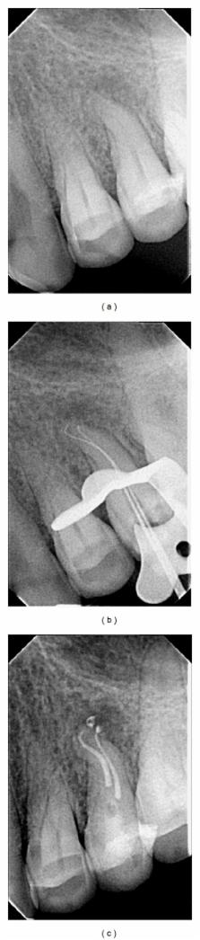
          Recognition of anatomical variations is a real challenge for clinicians undertaking therapy regardless of the teeth that are to be treated. The extent of the curvature is one of the most important variables that could lead to instrument fracture. In clinical conditions, two curves can be present in the same root canal trajectory. This type of geometry is denoted as the “S” shape, and it is a challenging condition. This report describes a different clinical and educational scenario where four specialists around the world present different approaches for the treatment of root canals with double curvatures or S-shaped canals. Endodontic therapy is a very nuanced and challenging science and art. The clinical and teaching experience of the authors show different approaches that can be successfully employed to treat challenging teeth having roots with multiple curves. The necessity of precise knowledge of the root canal morphology and its variation is also underlined.

          Related collections

          Most cited references28

          • Record: found
          • Abstract: not found
          • Article: not found

          The "balanced force" concept for instrumentation of curved canals.

            Bookmark
            • Record: found
            • Abstract: found
            • Article: not found

            Effects of root canal preparation on apical geometry assessed by micro-computed tomography.

            Previous micro-computed tomography analyses of root canal preparation provided data that were usually averaged over canal length. The aim of this study was to compare preparation effects on apical root canal geometry. Sixty extracted maxillary molars (180 canals) used in prior studies were reevaluated for analyses of the apical 4 mm. Teeth were scanned by using micro-computed tomography before and after canal shaping with FlexMaster, GT-Rotary, Lightspeed, ProFile, ProTaper, instruments or nickel-titanium K-files for hand instrumentation. Apical preparation was to a size #40 in mesiobuccal and distobuccal and #45 in palatal canals except for GT (#20) and ProTaper (#25 in mesiobuccal and distobuccal and #30 in palatal canals, respectively). Data for canal volume changes, the structure model index (quantifying canal cross sections), and untreated surface area were contrasted by using analysis of variance and Scheffé tests. Mean mesiobuccal, distobuccal, and palatal canal volumes increased after preparation (P < .05), but differences were noted for preparation techniques. GT rendered the smallest (0.20 +/- 0.14 mm(3)); K-files and ProFile showed the largest volume increases (0.51 +/- 0.20 mm(3) and 0.45 +/- 021 mm(3), P < .05). All canals were slightly rounder in the apical 4 mm after preparation indicated by nonsignificant increases in structure model index. Untreated areas ranged from 4%-100% and were larger in mesiobuccal and palatal canals than in distobuccal ones. Preparation with GT left significantly larger untreated areas in all canal types (P < .05); among root canal types, distobuccal canals had the least amounts of untreated surface areas. Apical canal geometry was affected differently by 6 preparation techniques; preparations with GT instruments to an apical size #20 left more canal surface untouched, which might affect the ability to disinfect root canals in maxillary molars.
              Bookmark
              • Record: found
              • Abstract: found
              • Article: not found

              Radiographic quality of root fillings performed by undergraduate students.

              To evaluate the radiographic technical quality of root fillings and the incidence of iatrogenic errors in treatment provided by undergraduate students. Endodontic records and periapical radiographs of 759 root filled teeth were selected following exclusion of 25 (3.3%). A final total of 734 cases were used to assess the technical quality of root fillings in 1109 root canals performed by 4(th) and 5(th) undergraduate students. Two variables were examined: the length and the density of the fillings, categorized as acceptable and non-acceptable. The presence of ledges, root perforation, foramen perforation and fractured instruments were also investigated. Chi-square tests were used for statistical analysis. Acceptable root fillings were found in 55% of canals. More ;acceptable' root fillings occurred in maxillary compared to mandibular teeth (58 and 51% respectively) (P < 0.05) and in anterior compared with premolar (71 and 61%) and molar root canals (39% respectively) (P < 0.05). Insignificantly more (5.4%) acceptable root fillings were provided by 5(th) year students. Most of the underfillings and iatrogenic errors were detected in molars for both academic years (53 and 65% respectively). Ledges were noted in 55% of cases treated by both academic years and significantly more foramen perforations were detected in the 5(th) compared with the 4(th) year students (P < 0.05). There is a need to improve the technical quality of root fillings performed by the undergraduate students, especially in molar teeth.
                Bookmark

                Author and article information

                Journal
                Case Rep Dent
                Case Rep Dent
                CRID
                Case Reports in Dentistry
                Hindawi Publishing Corporation
                2090-6447
                2090-6455
                2014
                16 July 2014
                : 2014
                : 359438
                Affiliations
                1University of São Paulo, Ribeirão Preto, Brazil
                2Private Practice Limited to Endodontics, Kalithea, Greece
                3University of Tlaxcala, Puebla, Mexico
                4Private Practice Limited to Endodontics, Monterrey, Mexico
                5Grande Rio University, Rio de Janeiro, Brazil
                Author notes

                Academic Editor: Juan José Segura-Egea

                Article
                10.1155/2014/359438
                4124786
                25143838
                096032a8-8be4-4274-b21c-51dd9760f1dd
                Copyright © 2014 Ricardo Machado et al.

                This is an open access article distributed under the Creative Commons Attribution License, which permits unrestricted use, distribution, and reproduction in any medium, provided the original work is properly cited.

                History
                : 18 February 2014
                : 21 June 2014
                : 21 June 2014
                Categories
                Case Report

                Dentistry
                Dentistry

                Comments

                Comment on this article