3
views
0
recommends
+1 Recommend
0 collections
    0
    shares
      • Record: found
      • Abstract: found
      • Article: found
      Is Open Access

      Extracellular Vesicle Molecular Signatures Characterize Metastatic Dynamicity in Ovarian Cancer

      research-article

      Read this article at

      Bookmark
          There is no author summary for this article yet. Authors can add summaries to their articles on ScienceOpen to make them more accessible to a non-specialist audience.

          Abstract

          Background

          Late-stage diagnosis of ovarian cancer, a disease that originates in the ovaries and spreads to the peritoneal cavity, lowers 5-year survival rate from 90% to 30%. Early screening tools that can: i) detect with high specificity and sensitivity before conventional tools such as transvaginal ultrasound and CA-125, ii) use non-invasive sampling methods and iii) longitudinally significantly increase survival rates in ovarian cancer are needed. Studies that employ blood-based screening tools using circulating tumor-cells, -DNA, and most recently tumor-derived small extracellular vesicles (sEVs) have shown promise in non-invasive detection of cancer before standard of care. Our findings in this study show the promise of a sEV-derived signature as a non-invasive longitudinal screening tool in ovarian cancer.

          Methods

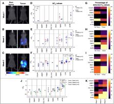
          Human serum samples as well as plasma and ascites from a mouse model of ovarian cancer were collected at various disease stages. Small extracellular vesicles (sEVs) were extracted using a commercially available kit. RNA was isolated from lysed sEVs, and quantitative RT-PCR was performed to identify specific metastatic gene expression.

          Conclusion

          This paper highlights the potential of sEVs in monitoring ovarian cancer progression and metastatic development. We identified a 7-gene panel in sEVs derived from plasma, serum, and ascites that overlapped with an established metastatic ovarian carcinoma signature. We found the 7-gene panel to be differentially expressed with tumor development and metastatic spread in a mouse model of ovarian cancer. The most notable finding was a significant change in the ascites-derived sEV gene signature that overlapped with that of the plasma-derived sEV signature at varying stages of disease progression. While there were quantifiable changes in genes from the 7-gene panel in serum-derived sEVs from ovarian cancer patients, we were unable to establish a definitive signature due to low sample number. Taken together our findings show that differential expression of metastatic genes derived from circulating sEVs present a minimally invasive screening tool for ovarian cancer detection and longitudinal monitoring of molecular changes associated with progression and metastatic spread.

          Related collections

          Most cited references74

          • Record: found
          • Abstract: found
          • Article: found
          Is Open Access

          Minimal information for studies of extracellular vesicles 2018 (MISEV2018): a position statement of the International Society for Extracellular Vesicles and update of the MISEV2014 guidelines

          ABSTRACT The last decade has seen a sharp increase in the number of scientific publications describing physiological and pathological functions of extracellular vesicles (EVs), a collective term covering various subtypes of cell-released, membranous structures, called exosomes, microvesicles, microparticles, ectosomes, oncosomes, apoptotic bodies, and many other names. However, specific issues arise when working with these entities, whose size and amount often make them difficult to obtain as relatively pure preparations, and to characterize properly. The International Society for Extracellular Vesicles (ISEV) proposed Minimal Information for Studies of Extracellular Vesicles (“MISEV”) guidelines for the field in 2014. We now update these “MISEV2014” guidelines based on evolution of the collective knowledge in the last four years. An important point to consider is that ascribing a specific function to EVs in general, or to subtypes of EVs, requires reporting of specific information beyond mere description of function in a crude, potentially contaminated, and heterogeneous preparation. For example, claims that exosomes are endowed with exquisite and specific activities remain difficult to support experimentally, given our still limited knowledge of their specific molecular machineries of biogenesis and release, as compared with other biophysically similar EVs. The MISEV2018 guidelines include tables and outlines of suggested protocols and steps to follow to document specific EV-associated functional activities. Finally, a checklist is provided with summaries of key points.
            Bookmark
            • Record: found
            • Abstract: found
            • Article: not found

            The biology, function, and biomedical applications of exosomes

            The study of extracellular vesicles (EVs) has the potential to identify unknown cellular and molecular mechanisms in intercellular communication and in organ homeostasis and disease. Exosomes, with an average diameter of ~100 nanometers, are a subset of EVs. The biogenesis of exosomes involves their origin in endosomes, and subsequent interactions with other intracellular vesicles and organelles generate the final content of the exosomes. Their diverse constituents include nucleic acids, proteins, lipids, amino acids, and metabolites, which can reflect their cell of origin. In various diseases, exosomes offer a window into altered cellular or tissue states, and their detection in biological fluids potentially offers a multicomponent diagnostic readout. The efficient exchange of cellular components through exosomes can inform their applied use in designing exosome-based therapeutics.
              Bookmark
              • Record: found
              • Abstract: not found
              • Article: not found

              Analyzing real-time PCR data by the comparative CT method

                Bookmark

                Author and article information

                Contributors
                Journal
                Front Oncol
                Front Oncol
                Front. Oncol.
                Frontiers in Oncology
                Frontiers Media S.A.
                2234-943X
                18 November 2021
                2021
                : 11
                : 718408
                Affiliations
                [1] 1 Department of Biomedical Engineering, Rutgers University , Piscataway, NJ, United States
                [2] 2 Department of Chemical and Biochemical Engineering, Rutgers University , Piscataway, NJ, United States
                [3] 3 Rutgers-Robert Wood Johnson Medical School, Rutgers University , New Brunswick, NJ, United States
                [4] 4 School of Environmental and Biological Sciences, Rutgers University , New Brunswick, NJ, United States
                [5] 5 Rutgers Cancer Institute of New Jersey, Rutgers University , New Brunswick, NJ, United States
                Author notes

                Edited by: Hao Zhang, Jinan University, China

                Reviewed by: Marcio Chaim Bajgelman, National Center for Research in Energy and Materials, Brazil; Dianzheng Zhang, Philadelphia College of Osteopathic Medicine (PCOM), United States

                *Correspondence: Vidya Ganapathy, vg180@ 123456soe.rutgers.edu ; Nicola L. Francis, nicola.francis@ 123456rutgers.edu

                This article was submitted to Molecular and Cellular Oncology, a section of the journal Frontiers in Oncology

                Article
                10.3389/fonc.2021.718408
                8637407
                08f3a520-61d0-4c12-8bb9-b5f2acc24609
                Copyright © 2021 Gonda, Zhao, Shah, Siebert, Gunda, Inan, Kwon, Libutti, Moghe, Francis and Ganapathy

                This is an open-access article distributed under the terms of the Creative Commons Attribution License (CC BY). The use, distribution or reproduction in other forums is permitted, provided the original author(s) and the copyright owner(s) are credited and that the original publication in this journal is cited, in accordance with accepted academic practice. No use, distribution or reproduction is permitted which does not comply with these terms.

                History
                : 31 May 2021
                : 29 October 2021
                Page count
                Figures: 6, Tables: 1, Equations: 0, References: 74, Pages: 14, Words: 8054
                Funding
                Funded by: National Institute of Biomedical Imaging and Bioengineering , doi 10.13039/100000070;
                Categories
                Oncology
                Original Research

                Oncology & Radiotherapy
                extracellular vesicle,exosome,gene signatures,metastasis,ovarian cancer 2
                Oncology & Radiotherapy
                extracellular vesicle, exosome, gene signatures, metastasis, ovarian cancer 2

                Comments

                Comment on this article