6
views
0
recommends
+1 Recommend
0 collections
    0
    shares
      • Record: found
      • Abstract: found
      • Article: found
      Is Open Access

      Growth Differentiation Factor 15 May Predict Mortality of Peripheral and Coronary Artery Diseases and Correlate with Their Risk Factors

      research-article

      Read this article at

      Bookmark
          There is no author summary for this article yet. Authors can add summaries to their articles on ScienceOpen to make them more accessible to a non-specialist audience.

          Abstract

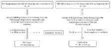
          Plasma GDF15 concentrations were measured in 612 Taiwanese individuals without overt systemic disease. Clinical parameters, GDF15 genetic variants, and 22 biomarker levels were analyzed. We further enrolled 86 patients with PAD and 481 patients with CAD, who received endovascular intervention and coronary angiography, respectively, to examine the role of GDF15 level in predicting all-cause mortality. Significant associations were found between GDF15 genotypes/haplotypes and GDF15 levels. The circulating GDF15 level was positively associated with age, smoking, hypertension, and diabetes mellitus as well as circulating levels of lipocalin 2 and various biomarkers of inflammation and oxidative stress. Kaplan-Meier survival analysis showed that baseline GDF15 levels of above 3096 pg/mL and 1123 pg/mL were strong predictors of death for patients with PAD and CAD, respectively ( P = 0.011 and P < 0.001). GDF15 more accurately reclassified 17.3% and 29.2% of patients with PAD and CAD, respectively ( P = 0.0046 and P = 0.0197), compared to C-reactive protein. Both genetic and nongenetic factors, including cardiometabolic and inflammatory markers and adipokines, were significantly associated with GDF15 level. A high level of GDF15 was significantly associated with an increase of all-cause mortality in patients with high-risk PAD and in patients with angiographically documented CAD.

          Related collections

          Most cited references43

          • Record: found
          • Abstract: found
          • Article: not found

          MIC-1, a novel macrophage inhibitory cytokine, is a divergent member of the TGF-beta superfamily.

          Macrophages play a key role in both normal and pathological processes involving immune and inflammatory responses, to a large extent through their capacity to secrete a wide range of biologically active molecules. To identify some of these as yet not characterized molecules, we have used a subtraction cloning approach designed to identify genes expressed in association with macrophage activation. One of these genes, designated macrophage inhibitory cytokine 1 (MIC-1), encodes a protein that bears the structural characteristics of a transforming growth factor beta (TGF-beta) superfamily cytokine. Although it belongs to this superfamily, it has no strong homology to existing families, indicating that it is a divergent member that may represent the first of a new family within this grouping. Expression of MIC-1 mRNA in monocytoid cells is up-regulated by a variety of stimuli associated with activation, including interleukin 1beta, tumor necrosis factor alpha (TNF-alpha), interleukin 2, and macrophage colony-stimulating factor but not interferon gamma, or lipopolysaccharide (LPS). Its expression is also increased by TGF-beta. Expression of MIC-1 in CHO cells results in the proteolytic cleavage of the propeptide and secretion of a cysteine-rich dimeric protein of Mr 25 kDa. Purified recombinant MIC-1 is able to inhibit lipopolysaccharide -induced macrophage TNF-alpha production, suggesting that MIC-1 acts in macrophages as an autocrine regulatory molecule. Its production in response to secreted proinflammatory cytokines and TGF-beta may serve to limit the later phases of macrophage activation.
            Bookmark
            • Record: found
            • Abstract: found
            • Article: not found

            The transforming growth factor-beta superfamily member growth-differentiation factor-15 protects the heart from ischemia/reperfusion injury.

            Data from the Women's Health Study show that serum levels of growth-differentiation factor-15 (GDF-15), a distant member of the transforming growth factor-beta superfamily, are an independent risk indicator for adverse cardiovascular events. However, the cellular sources, upstream regulators, and functional effects of GDF-15 in the cardiovascular system have not been elucidated. We have identified GDF-15 by cDNA expression array analysis as a gene that is strongly upregulated by nitrosative stress in cultured cardiomyocytes isolated from 1- to 3-day-old rats. GDF-15 mRNA and pro-peptide expression levels were also induced in cardiomyocytes subjected to simulated ischemia/reperfusion (I/R) via NO-peroxynitrite-dependent signaling pathways. GDF-15 was actively secreted into the culture supernatant, suggesting that it might exert autocrine/paracrine effects during I/R. To explore the in vivo relevance of these findings, mice were subjected to transient or permanent coronary artery ligation. Myocardial GDF-15 mRNA and pro-peptide abundance rapidly increased in the area-at-risk after ischemic injury. Similarly, patients with an acute myocardial infarction had enhanced myocardial GDF-15 pro-peptide expression levels. As shown by immunohistochemistry, cardiomyocytes in the ischemic area contributed significantly to the induction of GDF-15 in the infarcted human heart. To delineate the function of GDF-15 during I/R, Gdf-15 gene-targeted mice were subjected to transient coronary artery ligation for 1 hour followed by reperfusion for 24 hours. Gdf-15-deficient mice developed greater infarct sizes and displayed more cardiomyocyte apoptosis in the infarct border zone after I/R compared with wild-type littermates, indicating that endogenous GDF-15 limits myocardial tissue damage in vivo. Moreover, treatment with recombinant GDF-15 protected cultured cardiomyocytes from apoptosis during simulated I/R as shown by histone ELISA, TUNEL/Hoechst staining, and annexin V/propidium iodide fluorescence-activated cell sorting (FACS) analysis. Mechanistically, the prosurvival effects of GDF-15 in cultured cardiomyocytes were abolished by phosphoinositide 3-OH kinase inhibitors and adenoviral expression of dominant-negative Akt1 (K179M mutation). In conclusion, our study identifies induction of GDF-15 in the heart as a novel defense mechanism that protects from I/R injury.
              Bookmark
              • Record: found
              • Abstract: found
              • Article: not found

              GDF-15 is an inhibitor of leukocyte integrin activation required for survival after myocardial infarction in mice.

              Inflammatory cell recruitment after myocardial infarction needs to be tightly controlled to permit infarct healing while avoiding fatal complications such as cardiac rupture. Growth differentiation factor-15 (GDF-15), a transforming growth factor-β (TGF-β)-related cytokine, is induced in the infarcted heart of mice and humans. We show that coronary artery ligation in Gdf15-deficient mice led to enhanced recruitment of polymorphonuclear leukocytes (PMNs) into the infarcted myocardium and an increased incidence of cardiac rupture. Conversely, infusion of recombinant GDF-15 repressed PMN recruitment after myocardial infarction. In vitro, GDF-15 inhibited PMN adhesion, arrest under flow and transendothelial migration. Mechanistically, GDF-15 counteracted chemokine-triggered conformational activation and clustering of β(2) integrins on PMNs by activating the small GTPase Cdc42 and inhibiting activation of the small GTPase Rap1. Intravital microscopy in vivo in Gdf15-deficient mice showed that Gdf-15 is required to prevent excessive chemokine-activated leukocyte arrest on the endothelium. Genetic ablation of β(2) integrins in myeloid cells rescued the mortality of Gdf15-deficient mice after myocardial infarction. To our knowledge, GDF-15 is the first cytokine identified as an inhibitor of PMN recruitment by direct interference with chemokine signaling and integrin activation. Loss of this anti-inflammatory mechanism leads to fatal cardiac rupture after myocardial infarction.
                Bookmark

                Author and article information

                Journal
                Mediators Inflamm
                Mediators Inflamm
                MI
                Mediators of Inflammation
                Hindawi
                0962-9351
                1466-1861
                2017
                17 July 2017
                : 2017
                : 9398401
                Affiliations
                1Cardiovascular Division, Department of Internal Medicine, Chang Gung Memorial Hospital and Chang Gung University College of Medicine, Taoyuan 33305, Taiwan
                2Department of Life Science, Chinese Culture University, Taipei 11114, Taiwan
                3Cardiovascular Center and Division of Cardiology, Department of Internal Medicine, National Taiwan University Hospital and National Taiwan University College of Medicine, Taipei 10002, Taiwan
                4Department of Research, Taipei Tzu Chi Hospital, Buddhist Tzu Chi Medical Foundation, New Taipei City 23142, Taiwan
                5Cardiovascular Center and Division of Cardiology, Department of Internal Medicine, Taipei Tzu Chi Hospital, Buddhist Tzu Chi Medical Foundation, New Taipei City 23142, Taiwan
                6School of Medicine, Tzu Chi University, Hualien 97071, Taiwan
                Author notes

                Academic Editor: Mirella Giovarelli

                Author information
                http://orcid.org/0000-0001-7640-7702
                http://orcid.org/0000-0002-8218-6827
                Article
                10.1155/2017/9398401
                5535745
                28798540
                08449631-fc51-431b-a98d-050766512325
                Copyright © 2017 Lung-An Hsu et al.

                This is an open access article distributed under the Creative Commons Attribution License, which permits unrestricted use, distribution, and reproduction in any medium, provided the original work is properly cited.

                History
                : 11 March 2017
                : 13 June 2017
                Funding
                Funded by: Chang Gung Research Grant Foundation
                Award ID: CMRPG3E0871
                Funded by: National Science Council
                Award ID: NSC 101-2314-B-303 -023 -MY3
                Funded by: Tzu Chi University
                Award ID: TCIRP102001-02Y1
                Funded by: Buddhist Tzu Chi Medical Foundation
                Award ID: TCRD-TPE-106-C1-1
                Award ID: TCRD-TPE-106-RT-3
                Award ID: TCRD-TPE-103-RT-2
                Funded by: Taipei Tzu Chi Hospital
                Categories
                Research Article

                Immunology
                Immunology

                Comments

                Comment on this article