6
views
0
recommends
+1 Recommend
0 collections
    0
    shares
      • Record: found
      • Abstract: found
      • Article: found
      Is Open Access

      Human amniotic membrane plug to promote failed macular hole closure

      research-article

      Read this article at

      Bookmark
          There is no author summary for this article yet. Authors can add summaries to their articles on ScienceOpen to make them more accessible to a non-specialist audience.

          Abstract

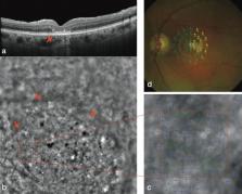
          The failed macular hole is a full-thickness defect involving the fovea that fails to close despite 1 or more surgeries. While many surgical options have been proposed to manage it, none of these guarantee complete anatomical success and satisfactory visual recovery. We report postoperative outcomes on 36 patients affected by failed macular hole, treated with a human amniotic membrane plug transplant. Follow-ups were performed with a standard ophthalmological examination and with advanced multimodal diagnostic imaging. Anatomical closure was achieved at 3 months in all patients. Mean best-corrected visual acuity improved statistically significantly at 6 months ( p < 0.05). Through microperimetric tests, we assessed a partial recovery of the macular sensitivity on the edges of the plug. Analyzing SD-OCT images, we reported a tissutal ingrowth above the plug, and its segmentation into layers, mimicking normal retinal architecture. OCT-Angiography images non invasively analysed the retinal parafoveal capillary microvasculature; the elaboration of Adaptive Optics images showed the presence of photoreceptors at the edges of the plug. This work demonstrates not only the complete anatomical success of our technique, but also remarkable functional results, and opens the door to a greater understanding of modifications induced by the presence of a human amniotic membrane plug.

          Related collections

          Most cited references27

          • Record: found
          • Abstract: found
          • Article: not found

          Inverted internal limiting membrane flap technique for large macular holes.

          Large macular holes usually have an increased risk of surgical failure. Up to 44% of large macular holes remain open after 1 surgery. Another 19% to 39% of macular holes are flat-open after surgery. Flat-open macular holes are associated with limited visual acuity. This article presents a modification of the standard macular hole surgery to improve functional and anatomic outcomes in patients with large macular holes. A prospective, randomized clinical trial. Patients with macular holes larger than 400 μm were included. In group 1, 51 eyes of 40 patients underwent standard 3-port pars plana vitrectomy with air. In group 2, 50 eyes of 46 patients underwent a modification of the standard technique, called the inverted internal limiting membrane (ILM) flap technique. In the inverted ILM flap technique, instead of completely removing the ILM after trypan blue staining, a remnant attached to the margins of the macular hole was left in place. This ILM remnant was then inverted upside-down to cover the macular hole. Fluid-air exchange was then performed. Spectral optical coherence tomography and clinical examination were performed before surgery and postoperatively at 1 week and 1, 3, 6, and 12 months. Visual acuity and postoperative macular hole closure. Preoperative mean visual acuity was 0.12 in group 1 and 0.078 in group 2. Macular hole closure was observed in 88% of patients in group 1 and in 98% of patients in group 2. A flat-hole roof with bare retinal pigment epithelium (flat-open) was observed in 19% of patients in group 1 and 2% of patients in group 2. Mean (or median) postoperative visual acuity 12 months after surgery was 0.17 (range, 0.1-0.6) in group 1 and 0.28 (range, 0.02-0.8) in group 2 (P = 0.001). The inverted ILM flap technique prevents the postoperative flat-open appearance of a macular hole and improves both the functional and anatomic outcomes of vitrectomy for macular holes with a diameter greater than 400 μm. Spectral optical coherence tomography after vitrectomy with the inverted ILM flap technique suggests improved foveal anatomy compared with the standard surgery. Copyright © 2010 American Academy of Ophthalmology. Published by Elsevier Inc. All rights reserved.
            Bookmark
            • Record: found
            • Abstract: found
            • Article: not found

            Autologous transplantation of the internal limiting membrane for refractory macular holes.

            To determine the effectiveness of autologous transplantation of the internal limiting membrane (ILM) for refractory macular holes.
              Bookmark
              • Record: found
              • Abstract: not found
              • Article: not found

              Autologous Neurosensory Retinal Free Flap for Closure of Refractory Myopic Macular Holes.

                Bookmark

                Author and article information

                Contributors
                tomaso.caporossi@gmail.com
                Journal
                Sci Rep
                Sci Rep
                Scientific Reports
                Nature Publishing Group UK (London )
                2045-2322
                26 October 2020
                26 October 2020
                2020
                : 10
                : 18264
                Affiliations
                [1 ]GRID grid.8404.8, ISNI 0000 0004 1757 2304, Department of NEUROFARBA, Ophthalmology, , University of Florence, ; Largo Brambilla 3, Careggi, 50134 Florence, Italy
                [2 ]Ospedale Oftalmico Di Torino, Turin, Italy
                [3 ]GRID grid.414603.4, Ophthalmology Unit, , Fondazione Policlinico Universitario A. Gemelli IRCCS, ; 00196 Rome, Italy
                [4 ]GRID grid.8142.f, ISNI 0000 0001 0941 3192, Department of Ophthalmology, , Catholic University of “Sacro Cuore”, ; 00168 Rome, Italy
                Article
                75292
                10.1038/s41598-020-75292-2
                7588413
                33106542
                0825be9d-7c62-4d05-843e-0c7849f265d1
                © The Author(s) 2020

                Open Access This article is licensed under a Creative Commons Attribution 4.0 International License, which permits use, sharing, adaptation, distribution and reproduction in any medium or format, as long as you give appropriate credit to the original author(s) and the source, provide a link to the Creative Commons licence, and indicate if changes were made. The images or other third party material in this article are included in the article's Creative Commons licence, unless indicated otherwise in a credit line to the material. If material is not included in the article's Creative Commons licence and your intended use is not permitted by statutory regulation or exceeds the permitted use, you will need to obtain permission directly from the copyright holder. To view a copy of this licence, visit http://creativecommons.org/licenses/by/4.0/.

                History
                : 8 June 2020
                : 22 September 2020
                Categories
                Article
                Custom metadata
                © The Author(s) 2020

                Uncategorized
                diseases,medical research
                Uncategorized
                diseases, medical research

                Comments

                Comment on this article