9
views
0
recommends
+1 Recommend
0 collections
    0
    shares
      • Record: found
      • Abstract: found
      • Article: not found

      Aggressive Angiomyxoma of the Vulva: A Bizarre Perineal Lesion

      case-report

      Read this article at

      ScienceOpenPublisherPMC
      Bookmark
          There is no author summary for this article yet. Authors can add summaries to their articles on ScienceOpen to make them more accessible to a non-specialist audience.

          Abstract

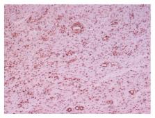
          Introduction. Aggressive angiomyxoma is a rare, slowly growing, and benign tumour of mesenchymal origin, which affects women of reproductive age and is associated with a high risk of local recurrence. Case Presentation. A case of a 47-year-old white female is presented herein, with a large polypoid, gelatinous mass on the right labia majora, measuring 26 × 21 × 6 cm. Histopathologically, the lesion was composed of spindle and stellate-shaped cells embedded in a myxoid matrix. Another specific feature was the presence of variable-sized thin-walled capillaries and thick-walled vascular channels. The patient underwent wide local excision of the tumour with clear margins and developed local recurrence 18 months later. Discussion. Aggressive angiomyxoma of the vulva needs to be distinguished from benign myxoid tumors with a low risk of local recurrence as well as from malignant myxoid neoplasms. Usually wide local excision with tumour-free margins and occasionally hormonal manipulation is the treatment of choice.

          Related collections

          Most cited references20

          • Record: found
          • Abstract: found
          • Article: not found

          Aggressive angiomyxoma of the female pelvis and perineum. Report of nine cases of a distinctive type of gynecologic soft-tissue neoplasm.

          Nine case of a distinctive soft tissue tumor of the female pelvis and perineum are described. They were characterized by their occurrence in young women (ages 21-38), large size (up to 60 X 20 cm), locally infiltrative nature, and grossly gelatinous appearance. The initial clinical impression was usually that of a Bartholin gland cyst. The microscopic appearance was that of spindle or stellate cells widely separated by a loose myxoid stroma focally rich in collagen fibrils, a prominent vascular component, including many large thick-walled vessels without an arborizing pattern, and foci of proliferating glandular elements in two cases. Mitotic activity was exceedingly low. Ultrastructural study of the spindle cells showed features consistent with myofibroblastic differentiation. Four patients developed large local recurrences; one tumor recurred twice, 14 and 15 years after initial excision. No distant metastases have been documented to date, and all patients are alive and well. The differential diagnosis of this unusual tumor includes myxoma, myxoid liposarcoma, sarcoma botryoides, myxoid variant of malignant fibrous histiocytoma, nerve sheath myxoma, and other soft tissue tumors with secondary myxoid changes. We have chosen the term "aggressive angiomyxoma" for this neoplasm to emphasize the neoplastic nature of the blood vessels and its locally infiltrative and recurrent nature.
            Bookmark
            • Record: found
            • Abstract: not found
            • Article: not found

            Aggressive angiomyxoma of the female pelvis and perineum

              Bookmark
              • Record: found
              • Abstract: found
              • Article: not found

              Aggressive angiomyxoma of the vulva: Dramatic response to gonadotropin-releasing hormone agonist therapy.

              Aggressive angiomyxoma (AA) is a vulvovaginal mesenchymal neoplasm with a marked tendency to local recurrence but which usually does not metastasise. Most cases exhibit positive immunohistochemical staining with oestrogen receptor (ER) and, or, progesterone receptor (PR). We report a case of AA which exhibited positive immunohistochemical staining with ER and in which radiological examination following resection showed extensive residual tumour. The patient was commenced on gonadotropin-releasing hormone (GnRH) agonist therapy which resulted in complete radiological remission with replacement by scar tissue. The patient is currently maintained on a GnRH agonist. The present case, together with several others reported in the literature, suggests that GnRH agonists may be of value in managing cases of AA, either primary or recurrent, which are not amenable to surgical excision. These agents may also be used to effect a reduction in size, so that more conservative surgery can be undertaken.
                Bookmark

                Author and article information

                Journal
                Case Rep Oncol Med
                Case Rep Oncol Med
                CRIONM
                Case Reports in Oncological Medicine
                Hindawi Publishing Corporation
                2090-6706
                2090-6714
                2015
                22 April 2015
                : 2015
                : 292304
                Affiliations
                1Pathology Department, “Tzaneion” General Hospital, 185 36 Piraeus, Greece
                22nd Department of Surgery, “Tzaneion” General Hospital, 185 36 Piraeus, Greece
                3Radiology & Interventional Radiology Department, “Tzaneion” General Hospital, 185 36 Piraeus, Greece
                42nd Department of Surgery, University Hospital “Aretaeion”, 115 28 Athens, Greece
                Author notes
                *Despoina Myoteri: dmyoteri@ 123456gmail.com

                Academic Editor: Francesca Micci

                Article
                10.1155/2015/292304
                4421102
                25977826
                06f43c25-edb8-4b5e-a6d3-cfc162f819ed
                Copyright © 2015 Adamantia Zizi-Sermpetzoglou et al.

                This is an open access article distributed under the Creative Commons Attribution License, which permits unrestricted use, distribution, and reproduction in any medium, provided the original work is properly cited.

                History
                : 28 February 2015
                : 15 April 2015
                Categories
                Case Report

                Oncology & Radiotherapy
                Oncology & Radiotherapy

                Comments

                Comment on this article