34
views
0
recommends
+1 Recommend
0 collections
    0
    shares
      • Record: found
      • Abstract: found
      • Article: found
      Is Open Access

      Additive Effect of Platelet Rich Fibrin with Coronally Advanced Flap Procedure in Root Coverage of Miller’s Class I and II Recession Defects—A PRISMA Compliant Systematic Review and Meta-Analysis

      review-article

      Read this article at

      Bookmark
          There is no author summary for this article yet. Authors can add summaries to their articles on ScienceOpen to make them more accessible to a non-specialist audience.

          Abstract

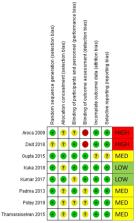
          Aim: This systematic review and meta-analysis aims to assess the additive effect of leukocyte and platelet-rich fibrin (L-PRF) on coronally advanced flap (CAF) procedures in root coverage of Miller’s class I and II gingival recession defects. Review methodology: A comprehensive search in MEDLINE (PubMed), Scopus and CENTRAL (the Cochrane Central Register of Controlled Trials), along with an additional hand search, provided eight randomized clinical trials to be included in this review. A total of 167 patients with 470 gingival recession defects were analyzed. A meta-analysis was carried out to assess the change in gingival thickness (GT), width of keratinized gingiva (WKG), root coverage percentage (%RC), clinical attachment level (CAL) and recession depth (RD) at all follow-ups between CAF alone and CAF + L-PRF groups for all included studies. A subgroup analysis was carried out based on recession type (single/multiple). Results: Overall, a significant improvement in GT, CAL and RD was found when treated with CAF + L-PRF. There was a trend for a positive effect in terms of an increase in WKG when using L-PRF, especially in the treatment of single recession, though significance was not achieved ( p = 0.08 overall). The results of heterogeneity among the subgroups were varied and were found to be greater than 91.3% for GT and 32.8% for WKG. Conclusion: L-PRF when used in addition to CAF showed favorable results for the treatment of class I and II gingival recession defects.

          Related collections

          Most cited references53

          • Record: found
          • Abstract: found
          • Article: not found

          Angiogenesis in wound healing.

          During wound healing, angiogenic capillary sprouts invade the fibrin/fibronectin-rich wound clot and within a few days organize into a microvascular network throughout the granulation tissue. As collagen accumulates in the granulation tissue to produce scar, the density of blood vessels diminishes. A dynamic interaction occurs among endothelial cells, angiogenic cytokines, such as FGF, VEGF, TGF-beta, angiopoietin, and mast cell tryptase, and the extracellular matrix (ECM) environment. Specific endothelial cell ECM receptors are critical for these morphogenetic changes in blood vessels during wound repair. In particular, alpha(v)beta3, the integrin receptor for fibrin and fibronectin, appears to be required for wound angiogenesis: alpha(v)beta3 is expressed on the tips of angiogenic capillary sprouts invading the wound clot, and functional inhibitors of alpha(v)beta3 transiently inhibit granulation tissue formation. Recent investigations have shown that the wound ECM can regulate angiogenesis in part by modulating integrin receptor expression. mRNA levels of alpha(v)beta3 in human dermal microvascular endothelial cells either plated on fibronectin or overlaid by fibrin gel were higher than in cells plated on collagen or overlaid by collagen gel. Wound angiogenesis also appears to be regulated by endothelial cell interaction with the specific three-dimensional ECM environment in the wound space. In an in vitro model of human sprout angiogenesis, three-dimensional fibrin gel, simulating early wound clot, but not collagen gel, simulating late granulation tissue, supported capillary sprout formation. Understanding the molecular mechanisms that regulate wound angiogenesis, particularly how ECM modulates ECM receptor and angiogenic factor requirements, may provide new approaches for treating chronic wounds.
            Bookmark
            • Record: found
            • Abstract: found
            • Article: not found

            Platelet-rich fibrin (PRF): a second-generation platelet concentrate. Part IV: clinical effects on tissue healing.

            Platelet-rich fibrin (PRF) belongs to a new generation of platelet concentrates, with simplified processing and without biochemical blood handling. In this fourth article, investigation is made into the previously evaluated biology of PRF with the first established clinical results, to determine the potential fields of application for this biomaterial. The reasoning is structured around 4 fundamental events of cicatrization, namely, angiogenesis, immune control, circulating stem cells trapping, and wound-covering epithelialization. All of the known clinical applications of PRF highlight an accelerated tissue cicatrization due to the development of effective neovascularization, accelerated wound closing with fast cicatricial tissue remodelling, and nearly total absence of infectious events. This initial research therefore makes it possible to plan several future PRF applications, including plastic and bone surgery, provided that the real effects are evaluated both impartially and rigorously.
              Bookmark
              • Record: found
              • Abstract: found
              • Article: not found

              Slow release of growth factors and thrombospondin-1 in Choukroun's platelet-rich fibrin (PRF): a gold standard to achieve for all surgical platelet concentrates technologies.

              Platelet concentrates for surgical topical applications are nowadays often used, but quantification of the long-term growth factor release from these preparations in most cases is impossible. Indeed, in most protocols, platelets are massively activated and there is no significant fibrin matrix to support growth factor release and cell migration. Choukroun's platelet-rich fibrin (PRF), a second generation platelet concentrate, is a leucocyte- and platelet-rich fibrin biomaterial. Here, we show that this dense fibrin membrane releases high quantities of three main growth factors (Transforming Growth Factor b-1 (TGFbeta-1), platelet derived growth factor AB, PDGF-AB; vascular endothelial growth factor, VEGF) and an important coagulation matricellular glycoprotein (thrombospondin-1, TSP-1) during 7 days. Moreover, the comparison between the final released amounts and the initial content of the membrane (after forcible extraction) allows us to consider that the leucocytes trapped in the fibrin matrix continue to produce high quantities of TGFbeta-1 and VEGF during the whole experimental time.
                Bookmark

                Author and article information

                Journal
                Materials (Basel)
                Materials (Basel)
                materials
                Materials
                MDPI
                1996-1944
                27 September 2020
                October 2020
                : 13
                : 19
                : 4314
                Affiliations
                [1 ]Department of Periodontics and Oral Implantology, Siksha ‘O’ Anusandhan University, Bhubaneswar 751003, India; sauravpanda@ 123456soa.ac.in (S.P.); anuragsatpathy@ 123456soa.ac.in (A.S.); abhayadas@ 123456soa.ac.in (A.C.D.); manojkumar@ 123456soa.ac.in (M.K.)
                [2 ]Department of Biomedical, Surgical and Dental Sciences, Università degli Studi di Milano, 20122 Milano, Italy; silvio.taschieri@ 123456unimi.it
                [3 ]Department of Conservative Dentistry and Endodontics, Institute of Dental Sciences, Siksha ‘O’ Anusandhan University, Bhubaneswar 751003, India; loramishra@ 123456soa.ac.in
                [4 ]Private Practice, Gainesville, FL 32603, USA; swati.gup9@ 123456gmail.com
                [5 ]Department of Prosthodontics, Institute of Dental Sciences, Siksha ‘O’ Anusandhan University, Bhubaneswar 751003, India; drgunjans22@ 123456gmail.com
                [6 ]Department of General Dentistry, Medical University of Lodz, 92-213 Lodz, Poland; monika.lukomska-szymanska@ 123456umed.lodz.pl
                [7 ]Dental Clinic, IRCCS Istituto Ortopedico Galeazzi, 20161 Milano, Italy
                Author notes
                [* ]Correspondence: massimo.delfabbro@ 123456unimi.it ; Tel.: +39-02-5031-9950; Fax: +39-02-5031-9960
                Author information
                https://orcid.org/0000-0002-5648-4281
                https://orcid.org/0000-0001-7832-3406
                https://orcid.org/0000-0002-8680-2172
                https://orcid.org/0000-0003-1088-2265
                https://orcid.org/0000-0002-6110-4298
                https://orcid.org/0000-0002-7866-5024
                https://orcid.org/0000-0001-7144-0984
                Article
                materials-13-04314
                10.3390/ma13194314
                7579296
                32992613
                05d4ca3a-b3f0-445b-93f3-7645fdefbb6b
                © 2020 by the authors.

                Licensee MDPI, Basel, Switzerland. This article is an open access article distributed under the terms and conditions of the Creative Commons Attribution (CC BY) license ( http://creativecommons.org/licenses/by/4.0/).

                History
                : 25 August 2020
                : 24 September 2020
                Categories
                Review

                gingival recession,periodontal disease,periodontal regeneration,blood platelet

                Comments

                Comment on this article