36
views
0
recommends
+1 Recommend
0 collections
    0
    shares
      • Record: found
      • Abstract: found
      • Article: found
      Is Open Access

      Analysis of the genomic landscape of multiple myeloma highlights novel prognostic markers and disease subgroups

      research-article

      Read this article at

      Bookmark
          There is no author summary for this article yet. Authors can add summaries to their articles on ScienceOpen to make them more accessible to a non-specialist audience.

          Abstract

          In multiple myeloma, next-generation sequencing (NGS) has expanded our knowledge of genomic lesions, and highlighted a dynamic and heterogeneous composition of the tumor. Here we used NGS to characterize the genomic landscape of 418 multiple myeloma cases at diagnosis and correlate this with prognosis and classification. Translocations and copy number abnormalities (CNAs) had a preponderant contribution over gene mutations in defining the genotype and prognosis of each case. Known and novel independent prognostic markers were identified in our cohort of proteasome inhibitor and immunomodulatory drug-treated patients with long follow-up, including events with context-specific prognostic value, such as deletions of the PRDM1 gene. Taking advantage of the comprehensive genomic annotation of each case, we used innovative statistical approaches to identify potential novel myeloma subgroups. We observed clusters of patients stratified based on the overall number of mutations and number/type of CNAs, with distinct effects on survival, suggesting that extended genotype of multiple myeloma at diagnosis may lead to improved disease classification and prognostication.

          Related collections

          Most cited references27

          • Record: found
          • Abstract: found
          • Article: not found

          Genomic complexity of multiple myeloma and its clinical implications

          In the past 5 years, results from large-scale whole-exome sequencing studies have brought new insight into the clonal heterogeneity and evolution of multiple myeloma, a genetically complex disease. Herein, the authors describe the driver gene alterations and sequential acquisition of the main genomic aberrations involved in this disease, with a focus on the clonal heterogeneity of multiple myeloma and its clinical implications.
            Bookmark
            • Record: found
            • Abstract: found
            • Article: found
            Is Open Access

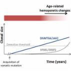
            Leukemia-Associated Somatic Mutations Drive Distinct Patterns of Age-Related Clonal Hemopoiesis

            Summary Clonal hemopoiesis driven by leukemia-associated gene mutations can occur without evidence of a blood disorder. To investigate this phenomenon, we interrogated 15 mutation hot spots in blood DNA from 4,219 individuals using ultra-deep sequencing. Using only the hot spots studied, we identified clonal hemopoiesis in 0.8% of individuals under 60, rising to 19.5% of those ≥90 years, thus predicting that clonal hemopoiesis is much more prevalent than previously realized. DNMT3A-R882 mutations were most common and, although their prevalence increased with age, were found in individuals as young as 25 years. By contrast, mutations affecting spliceosome genes SF3B1 and SRSF2, closely associated with the myelodysplastic syndromes, were identified only in those aged >70 years, with several individuals harboring more than one such mutation. This indicates that spliceosome gene mutations drive clonal expansion under selection pressures particular to the aging hemopoietic system and explains the high incidence of clonal disorders associated with these mutations in advanced old age.
              Bookmark
              • Record: found
              • Abstract: found
              • Article: not found

              A novel prognostic model in myeloma based on co-segregating adverse FISH lesions and the ISS: analysis of patients treated in the MRC Myeloma IX trial

              The association of genetic lesions detected by FISH with survival was analyzed in 1069 patients with newly presenting myeloma treated in the Medical Research Council (MRC) Myeloma IX trial, with the aim of identifying patients associated with the worst prognosis. A comprehensive FISH panel was performed, and the lesions associated with short PFS and OS in multivariate analysis were +1q21, del(17p13) and an adverse IGH translocation group incorporating t(4;14), t(14;16) and t(14;20). These lesions frequently co-segregated, and there was an association between the accumulation of these adverse FISH lesions and a progressive impairment of survival. This observation was used to define a series of risk groups based on number of adverse lesions. Taking this approach we defined a favorable risk group by the absence of adverse genetic lesions, an intermediate group with 1 adverse lesion and a high risk group defined by the co-segregation of >1 adverse lesion. This genetic grouping was independent of the ISS and so was integrated with the ISS to identify an ultra-high risk group defined by ISS II or III and >1 adverse lesion. This group constituted 13.8 % of patients and was associated with a median OS of 19.4 months.
                Bookmark

                Author and article information

                Contributors
                1 617 632 5607 , Nikhil_Munshi@dfci.harvard.edu
                Journal
                Leukemia
                Leukemia
                Leukemia
                Nature Publishing Group UK (London )
                0887-6924
                1476-5551
                22 May 2018
                22 May 2018
                2018
                : 32
                : 12
                : 2604-2616
                Affiliations
                [1 ]ISNI 0000 0004 1757 2822, GRID grid.4708.b, Department of Oncology and Onco-Hematology, , University of Milan, ; Milan, Italy
                [2 ]ISNI 0000 0001 0807 2568, GRID grid.417893.0, Department of Medical Oncology and Hematology, , Fondazione IRCCS Istituto Nazionale dei Tumori, ; Milan, Italy
                [3 ]ISNI 0000 0004 0606 5382, GRID grid.10306.34, Cancer Genome Project, , Wellcome Trust Sanger Institute, ; Cambridge, UK
                [4 ]ISNI 0000 0001 2171 9952, GRID grid.51462.34, Department of Epidemiology and Biostatistics, , Memorial Sloan Kettering Cancer Center, ; New York, NY USA
                [5 ]ISNI 0000 0001 2106 9910, GRID grid.65499.37, Harvard Medical School, LeBow Institute for Myeloma Therapeutics and Jerome Lipper Center for Multiple Myeloma Research, , Dana-Farber Cancer Institute, ; Boston, MA USA
                [6 ]European Bioinformatics Institute, Computational and Cancer Biology, Cambridge, UK
                [7 ]ISNI 0000 0001 2155 0800, GRID grid.5216.0, Department of Clinical Therapeutics, , National and Kapodistrian University of Athens, ; Athens, Greece
                [8 ]ISNI 0000 0004 0472 0371, GRID grid.277151.7, Department of Hematology, , University Hospital Hôtel-Dieu, ; Nantes, France
                [9 ]GRID grid.4817.a, CRCINA, INSERM, CNRS, Université d’Angers, , Université de Nantes, ; Nantes, France
                [10 ]Institute Universitaire du Cancer de Toulouse Oncopole, Toulouse, France
                [11 ]ISNI 0000 0001 1457 2980, GRID grid.411175.7, University Hospital, ; Toulouse, France
                Author information
                http://orcid.org/0000-0002-1018-5139
                http://orcid.org/0000-0002-5634-1539
                Article
                37
                10.1038/s41375-018-0037-9
                6286326
                29789651
                05d44df2-f516-4d8e-b335-ac447f77e8db
                © The Author(s) 2018

                Open Access This article is licensed under a Creative Commons Attribution-NonCommercial-NoDerivatives 4.0 International License, which permits any non-commercial use, sharing, distribution and reproduction in any medium or format, as long as you give appropriate credit to the original author(s) and the source, and provide a link to the Creative Commons license. You do not have permission under this license to share adapted material derived from this article or parts of it. The images or other third party material in this article are included in the article’s Creative Commons license, unless indicated otherwise in a credit line to the material. If material is not included in the article’s Creative Commons license and your intended use is not permitted by statutory regulation or exceeds the permitted use, you will need to obtain permission directly from the copyright holder. To view a copy of this license, visit http://creativecommons.org/licenses/by-nc-nd/4.0/.

                History
                : 18 August 2017
                : 28 October 2017
                : 10 November 2017
                Categories
                Article
                Custom metadata
                © Springer Nature Limited 2018

                Oncology & Radiotherapy
                Oncology & Radiotherapy

                Comments

                Comment on this article