6
views
0
recommends
+1 Recommend
0 collections
    0
    shares
      • Record: found
      • Abstract: found
      • Article: found
      Is Open Access

      Designing a large field-of-view two-photon microscope using optical invariant analysis

      research-article

      Read this article at

      Bookmark
          There is no author summary for this article yet. Authors can add summaries to their articles on ScienceOpen to make them more accessible to a non-specialist audience.

          Abstract.

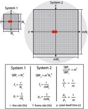
          Conventional two-photon microscopy (TPM) is capable of imaging neural dynamics with subcellular resolution, but it is limited to a field-of-view (FOV) diameter < 1    mm . Although there has been recent progress in extending the FOV in TPM, a principled design approach for developing large FOV TPM (LF-TPM) with off-the-shelf components has yet to be established. Therefore, we present a design strategy that depends on analyzing the optical invariant of commercially available objectives, relay lenses, mirror scanners, and emission collection systems in isolation. Components are then selected to maximize the space-bandwidth product of the integrated microscope. In comparison with other LF-TPM systems, our strategy simplifies the sequence of design decisions and is applicable to extending the FOV in any microscope with an optical relay. The microscope we constructed with this design approach can image < 1.7 - μ m lateral and < 28 - μ m axial resolution over a 7-mm diameter FOV, which is a 100-fold increase in FOV compared with conventional TPM. As a demonstration of the potential that LF-TPM has on understanding the microarchitecture of the mouse brain across interhemispheric regions, we performed in vivo imaging of both the cerebral vasculature and microglia cell bodies over the mouse cortex.

          Related collections

          Most cited references36

          • Record: found
          • Abstract: found
          • Article: not found

          Functional imaging with cellular resolution reveals precise micro-architecture in visual cortex.

          Neurons in the cerebral cortex are organized into anatomical columns, with ensembles of cells arranged from the surface to the white matter. Within a column, neurons often share functional properties, such as selectivity for stimulus orientation; columns with distinct properties, such as different preferred orientations, tile the cortical surface in orderly patterns. This functional architecture was discovered with the relatively sparse sampling of microelectrode recordings. Optical imaging of membrane voltage or metabolic activity elucidated the overall geometry of functional maps, but is averaged over many cells (resolution >100 microm). Consequently, the purity of functional domains and the precision of the borders between them could not be resolved. Here, we labelled thousands of neurons of the visual cortex with a calcium-sensitive indicator in vivo. We then imaged the activity of neuronal populations at single-cell resolution with two-photon microscopy up to a depth of 400 microm. In rat primary visual cortex, neurons had robust orientation selectivity but there was no discernible local structure; neighbouring neurons often responded to different orientations. In area 18 of cat visual cortex, functional maps were organized at a fine scale. Neurons with opposite preferences for stimulus direction were segregated with extraordinary spatial precision in three dimensions, with columnar borders one to two cells wide. These results indicate that cortical maps can be built with single-cell precision.
            Bookmark
            • Record: found
            • Abstract: found
            • Article: not found

            Retinal waves coordinate patterned activity throughout the developing visual system.

            The morphological and functional development of the vertebrate nervous system is initially governed by genetic factors and subsequently refined by neuronal activity. However, fundamental features of the nervous system emerge before sensory experience is possible. Thus, activity-dependent development occurring before the onset of experience must be driven by spontaneous activity, but the origin and nature of activity in vivo remains largely untested. Here we use optical methods to show in live neonatal mice that waves of spontaneous retinal activity are present and propagate throughout the entire visual system before eye opening. This patterned activity encompassed the visual field, relied on cholinergic neurotransmission, preferentially initiated in the binocular retina and exhibited spatiotemporal correlations between the two hemispheres. Retinal waves were the primary source of activity in the midbrain and primary visual cortex, but only modulated ongoing activity in secondary visual areas. Thus, spontaneous retinal activity is transmitted through the entire visual system and carries patterned information capable of guiding the activity-dependent development of complex intra- and inter-hemispheric circuits before the onset of vision.
              Bookmark
              • Record: found
              • Abstract: found
              • Article: not found

              A Cellular Resolution Map of Barrel Cortex Activity during Tactile Behavior.

              Comprehensive measurement of neural activity remains challenging due to the large numbers of neurons in each brain area. We used volumetric two-photon imaging in mice expressing GCaMP6s and nuclear red fluorescent proteins to sample activity in 75% of superficial barrel cortex neurons across the relevant cortical columns, approximately 12,000 neurons per animal, during performance of a single whisker object localization task. Task-related activity peaked during object palpation. An encoding model related activity to behavioral variables. In the column corresponding to the spared whisker, 300 layer (L) 2/3 pyramidal neurons (17%) each encoded touch and whisker movements. Touch representation declined by half in surrounding columns; whisker movement representation was unchanged. Following the emergence of stereotyped task-related movement, sensory representations showed no measurable plasticity. Touch direction was topographically organized, with distinct organization for passive and active touch. Our work reveals sparse and spatially intermingled representations of multiple tactile features. VIDEO ABSTRACT.
                Bookmark

                Author and article information

                Journal
                Neurophotonics
                Neurophotonics
                NEUROW
                NPh
                Neurophotonics
                Society of Photo-Optical Instrumentation Engineers
                2329-423X
                2329-4248
                19 February 2018
                April 2018
                : 5
                : 2
                : 025001
                Affiliations
                [a ]Washington University in Saint Louis , Department of Biomedical Engineering, St. Louis, Missouri, United States
                [b ]Washington University School of Medicine , Department of Radiology, St. Louis, Missouri, United States
                [c ]Washington University in Saint Louis , Department of Biology, St. Louis, Missouri, United States
                [d ]Washington University School of Medicine , Department of Neurology, St. Louis, Missouri, United States
                [e ]Washington University in Saint Louis , Department of Physics, St. Louis, Missouri, United States
                [f ]Université Laval , Génie Physique et Optique, Département de Physique, Ville de Québec, Quebec, Canada
                Author notes
                [* ]Address all correspondence to: Joseph P. Culver, E-mail: culverj@ 123456wustl.edu
                Author information
                https://orcid.org/0000-0002-3991-5879
                https://orcid.org/0000-0002-2852-8988
                https://orcid.org/0000-0001-6440-6948
                Article
                NPh-17125R 17125R
                10.1117/1.NPh.5.2.025001
                5818100
                29487876
                0589946e-cf70-4c90-8937-b8ed552050cc
                © The Authors.

                Published by SPIE under a Creative Commons Attribution 3.0 Unported License. Distribution or reproduction of this work in whole or in part requires full attribution of the original publication, including its DOI.

                History
                : 25 October 2017
                : 22 January 2018
                Page count
                Figures: 12, Tables: 6, References: 42, Pages: 20
                Funding
                Funded by: National Institutes of Health https://doi/10.13039/100000002
                Award ID: 1R01NS099429-01A1
                Award ID: 4R01NS078223-05
                Award ID: 5T32EB014855
                Funded by: McDonnell Center for Systems Neuroscience https://doi/10.13039/100009607
                Categories
                Research Papers
                Paper
                Custom metadata
                Bumstead et al.: Designing a large field-of-view two-photon microscope using optical invariant analysis

                two-photon microscopy,scanning microscopy,optical design,optical invariant,etendue,neurophysiology

                Comments

                Comment on this article