11
views
0
recommends
+1 Recommend
0 collections
    0
    shares
      • Record: found
      • Abstract: found
      • Article: found
      Is Open Access

      High expression ITGA2 affects the expression of MET, PD-L1, CD4 and CD8 with the immune microenvironment in pancreatic cancer patients

      research-article

      Read this article at

      Bookmark
          There is no author summary for this article yet. Authors can add summaries to their articles on ScienceOpen to make them more accessible to a non-specialist audience.

          Abstract

          Purpose

          Pancreatic cancer is characterized by a grim prognosis and is regarded as one of the most formidable malignancies. Among the genes exhibiting high expression in different tumor tissues, ITGA2 stands out as a promising candidate for cancer therapy. The promotion of cancer in pancreatic cancer is not effective. The objective of this study is to assess the presence of ITGA2, EMT and PD-L1 in pancreatic cancer.

          Experimental design

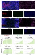
          We examined the expression of ITGA2, MET, E-cadherin, PD-L1, CD4, and CD8 proteins in 62 pancreatic cancer tissue samples using multi-tissue immunofluorescence and immunohistochemistry techniques. Functional assays, such as the cell migration assay and transwell assay, were used to determine the biological role of ITGA2 in pancreatic cancer. The relationship of ITGA2,EMT and PD-L1 were examined using Western blot analysis and RT-qPCR assay.

          Results

          In our study, we observed the expression of ITGA2, E-cadherin, and PD-L1 in both tumor and stroma tissues of pancreatic cancer. Additionally, a positive correlation between ITGA2, E-cadherin, and PD-L1 in the tumor region (r=0.559, P<0.001 and r=0.511, P<0.001), and PD-L1 in the stroma region (r=0.512, P<0.001).The expression levels of ITGA2, CD4, and CD8 were found to be higher in pancreatic cancer tissues compared to adjacent tissues (P < 0.05). Additionally, ITGA2 was negatively correlated with CD4 and CD8 (r = -0.344, P < 0.005 and r = -0.398, P < 0.005).Furthermore, ITGA2, CD4, and CD8 were found to be correlated with the survival time of patients (P < 0.05). Blocking ITGA2 inhibited the proliferation and invasion ability of pancreatic cancer cells significantly, Additionally, sh-ITGA2 can down-regulate the expression of EMT and PD-L1.

          Conclusions

          We identified a novel mechanism in which ITGA2 plays a crucial role in the regulation of pancreatic cancer growth and invasion. This mechanism involves the upregulation of MET and PD-L1 expression in pancreatic cancer cells. Additionally, we found that increased expression of ITGA2 is associated with a poor prognosis in pancreatic cancer patients. Furthermore, ITGA2 also affects immune regulation in these patients. Therefore, targeting ITGA2 is an effective method to enhance the efficacy of checkpoint immunotherapy and prohibiting tumor growth against pancreatic cancer.

          Related collections

          Most cited references37

          • Record: found
          • Abstract: found
          • Article: not found

          Analysis of relative gene expression data using real-time quantitative PCR and the 2(-Delta Delta C(T)) Method.

          The two most commonly used methods to analyze data from real-time, quantitative PCR experiments are absolute quantification and relative quantification. Absolute quantification determines the input copy number, usually by relating the PCR signal to a standard curve. Relative quantification relates the PCR signal of the target transcript in a treatment group to that of another sample such as an untreated control. The 2(-Delta Delta C(T)) method is a convenient way to analyze the relative changes in gene expression from real-time quantitative PCR experiments. The purpose of this report is to present the derivation, assumptions, and applications of the 2(-Delta Delta C(T)) method. In addition, we present the derivation and applications of two variations of the 2(-Delta Delta C(T)) method that may be useful in the analysis of real-time, quantitative PCR data. Copyright 2001 Elsevier Science (USA).
            Bookmark
            • Record: found
            • Abstract: found
            • Article: not found

            Cancer statistics, 2018

            Each year, the American Cancer Society estimates the numbers of new cancer cases and deaths that will occur in the United States and compiles the most recent data on cancer incidence, mortality, and survival. Incidence data, available through 2014, were collected by the Surveillance, Epidemiology, and End Results Program; the National Program of Cancer Registries; and the North American Association of Central Cancer Registries. Mortality data, available through 2015, were collected by the National Center for Health Statistics. In 2018, 1,735,350 new cancer cases and 609,640 cancer deaths are projected to occur in the United States. Over the past decade of data, the cancer incidence rate (2005-2014) was stable in women and declined by approximately 2% annually in men, while the cancer death rate (2006-2015) declined by about 1.5% annually in both men and women. The combined cancer death rate dropped continuously from 1991 to 2015 by a total of 26%, translating to approximately 2,378,600 fewer cancer deaths than would have been expected if death rates had remained at their peak. Of the 10 leading causes of death, only cancer declined from 2014 to 2015. In 2015, the cancer death rate was 14% higher in non-Hispanic blacks (NHBs) than non-Hispanic whites (NHWs) overall (death rate ratio [DRR], 1.14; 95% confidence interval [95% CI], 1.13-1.15), but the racial disparity was much larger for individuals aged <65 years (DRR, 1.31; 95% CI, 1.29-1.32) compared with those aged ≥65 years (DRR, 1.07; 95% CI, 1.06-1.09) and varied substantially by state. For example, the cancer death rate was lower in NHBs than NHWs in Massachusetts for all ages and in New York for individuals aged ≥65 years, whereas for those aged <65 years, it was 3 times higher in NHBs in the District of Columbia (DRR, 2.89; 95% CI, 2.16-3.91) and about 50% higher in Wisconsin (DRR, 1.78; 95% CI, 1.56-2.02), Kansas (DRR, 1.51; 95% CI, 1.25-1.81), Louisiana (DRR, 1.49; 95% CI, 1.38-1.60), Illinois (DRR, 1.48; 95% CI, 1.39-1.57), and California (DRR, 1.45; 95% CI, 1.38-1.54). Larger racial inequalities in young and middle-aged adults probably partly reflect less access to high-quality health care. CA Cancer J Clin 2018;68:7-30. © 2018 American Cancer Society.
              Bookmark
              • Record: found
              • Abstract: found
              • Article: not found

              Understanding the tumor immune microenvironment (TIME) for effective therapy

              The clinical successes in immunotherapy have been both astounding and at the same time unsatisfactory. Countless patients with varied tumor types have seen pronounced clinical response with immunotherapeutic intervention; however, many more patients have experienced minimal or no clinical benefit when provided the same treatment. As technology has advanced, so has the understanding of the complexity and diversity of the immune context of the tumor microenvironment and its influence on response to therapy. It has been possible to identify different subclasses of immune environment that have an influence on tumor initiation and response and therapy; by parsing the unique classes and subclasses of tumor immune microenvironment (TIME) that exist within a patient’s tumor, the ability to predict and guide immunotherapeutic responsiveness will improve, and new therapeutic targets will be revealed.
                Bookmark

                Author and article information

                Contributors
                Journal
                Front Immunol
                Front Immunol
                Front. Immunol.
                Frontiers in Immunology
                Frontiers Media S.A.
                1664-3224
                10 October 2023
                2023
                : 14
                : 1209367
                Affiliations
                [1] 1 1St Department of General Surgery, The First Affiliated Hospital of Dali University , Dali, Yunnan, China
                [2] 2 Clinical Medical College of Dali University , Dali, Yunnan, China
                Author notes

                Edited by: Qinyu Ge, Southeast University, China

                Reviewed by: Ling Zhi Wu, Nanjing University of Posts and Telecommunications, China; Ying Zhou, Southeast University, China

                *Correspondence: Yiming Chen, drchenyiming@ 123456gmail.com ; Yunbo Tan, tanyunbo6365z@ 123456163.com
                Article
                10.3389/fimmu.2023.1209367
                10594995
                37881431
                053f4532-75d4-4ada-b1c1-f0fc365b8b2a
                Copyright © 2023 Jin, Duan, Li, Li, Hu, Shi, Su, Li, Du, Chen and Tan

                This is an open-access article distributed under the terms of the Creative Commons Attribution License (CC BY). The use, distribution or reproduction in other forums is permitted, provided the original author(s) and the copyright owner(s) are credited and that the original publication in this journal is cited, in accordance with accepted academic practice. No use, distribution or reproduction is permitted which does not comply with these terms.

                History
                : 20 April 2023
                : 22 September 2023
                Page count
                Figures: 6, Tables: 3, Equations: 0, References: 37, Pages: 17, Words: 8964
                Funding
                This work is supported by Yunnan Provincial Foundation Joint Program for Local Undergraduate Universities, (202101BA070001-276); the Scientific Research Fund project of Yunnan Education Department (2021J0385).
                Categories
                Immunology
                Original Research
                Custom metadata
                Cancer Immunity and Immunotherapy

                Immunology
                pancreatic cancer,itga2,emt,pd-l1,microenvironment
                Immunology
                pancreatic cancer, itga2, emt, pd-l1, microenvironment

                Comments

                Comment on this article