- Record: found
- Abstract: found
- Article: not found
Immunoaffinity biosensors for the detection of SARS-CoV-1 using screened Fv-antibodies from an autodisplayed Fv-antibody library
Read this article at

Abstract
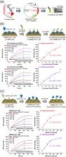
The detection of severe acute respiratory syndrome coronavirus (SARS-CoV-1) was demonstrated using screened Fv-antibodies for SPR biosensor and impedance spectrometry. The Fv-antibody library was first prepared on the outer membrane of E. coli using autodisplay technology and the Fv-variants (clones) with a specific affinity toward the SARS-CoV-1 spike protein (SP) were screened using magnetic beads immobilized with the SP. Upon screening the Fv-antibody library, two target Fv-variants (clones) with a specific binding affinity toward the SARS-CoV-1 SP were determined and the Fv-antibodies on two clones were named “Anti-SP1” (with CDR3 amino acid sequence: 1GRTTG 5NDRPD 11Y) and “Anti-SP2” (with CDR3 amino acid sequence: 1CLRQA 5GTADD 11V). The binding affinities of the two screened Fv-variants (clones) were analyzed using flow cytometry and the binding constants (K D) were estimated to be 80.5 ± 3.6 nM for Anti-SP1 and 45.6 ± 8.9 nM for Anti-SP2 (n = 3). In addition, the Fv-antibody including three CDR regions (CDR1, CDR2, and CDR3) and frame regions (FRs) between the CDR regions was expressed as a fusion protein (Mw. 40.6 kDa) with a green fluorescent protein (GFP) and the K D values of the expressed Fv-antibodies toward the SP estimated to be 15.3 ± 1.5 nM for Anti-SP1 (n = 3) and 16.3 ± 1.7 nM for Anti-SP2 (n = 3). Finally, the expressed Fv-antibodies screened against SARS-CoV-1 SP (Anti-SP1 and Anti-SP2) were applied for the detection of SARS-CoV-1. Consequently, the detection of SARS-CoV-1 was demonstrated to be feasible using the SPR biosensor and impedance spectrometry utilizing the immobilized Fv-antibodies against the SARS-CoV-1 SP.
Related collections
Most cited references76

- Record: found
- Abstract: found
- Article: found
Cryo-EM structure of the 2019-nCoV spike in the prefusion conformation
- Record: found
- Abstract: found
- Article: not found
Structure, Function, and Antigenicity of the SARS-CoV-2 Spike Glycoprotein
- Record: found
- Abstract: found
- Article: not found