243
views
0
recommends
+1 Recommend
1 collections
    0
    shares

      To submit to this journal, click here

      • Record: found
      • Abstract: found
      • Article: found
      Is Open Access

      Paragangliome malin orbitaire, à propos d’un cas Translated title: Malignant paraganglioma of the orbit, a case report

      case-report

      Read this article at

      Bookmark
          There is no author summary for this article yet. Authors can add summaries to their articles on ScienceOpen to make them more accessible to a non-specialist audience.

          Abstract

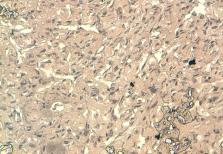
          Les paragangliomes sont des tumeurs neuroendocrines développées aux dépens du système nerveux parasympathique. Ils peuvent se localiser n’ importe où dans l’organisme depuis la tète et cou jusqu’au pelvis. La localisation orbitaire de cette tumeur est très rare. Nous présentons le cas d’un patient âgé de 37 ans qui présente depuis 4 mois une exophtalmie unilatérale droite, d’installation progressive, sans douleur ni baisse de l’acuité visuelle associés. L’examen général montre une tuméfaction sous le cuire chevelu, sans adénopathies locorégionales ni hépato ou splénomégalie. La tomodensitométrie retrouve un processus tumoral occupant le cadran supéro-externe de l’orbite droite, mesurant 38 mm de grand axe, envahissant la paroi supérieure et externe de l’orbite avec une importante ostéolyse. Un body scan révèle alors une métastase pulmonaire. L’examen histopathologique complétés par l’immunohistochimie, réalisé après biopsie, révèle un marquage cytoplasmique par l’anticorps anti-chromogranin, l’anticorps anti-synaptophysine et un marquage des vaisseaux par l’anticorps anti-CD31 soulignant l’architecture en zellbalen des nids tumoraux. Cet aspect est en faveur d’un paragangliome malin. Une exérèse chirurgicale incomplète suivie d’une radiothérapie adjuvante, sont alors réalisés. L’origine exacte de cette tumeur au sein de l’orbite reste très controversée. L’exophtalmie reste le principal signe révélateur. La tomodensitométrie, l’imagerie par résonnance magnétique et la scintigraphie au Metaiodobenzylguanidine radioinonisée à l’iode (MIBG-I 131) permettent d’orienter le diagnostic et faire un bilan d’extension de la tumeur. Le diagnostic de certitude repose sur l’histopathologie et l’immunohistochimie. L’excision totale de la lésion est le traitement de choix pour les lésions bien délimitées. Dans les formes plus étendues le traitement repose sur l’excision incomplète associée à une radiothérapie adjuvante ou au MIBG I 131. La localisation orbitaire du paragangliome reste très rare. Son diagnostic est difficile et repose essentiellement sur l’immunohistochimie. Son pronostic dépend essentiellement de l’extension locale et de la présence de métastases à distance qui signe le caractère malin du paragangliome.

          Most cited references11

          • Record: found
          • Abstract: found
          • Article: not found

          Phase II study of high-dose [131I]metaiodobenzylguanidine therapy for patients with metastatic pheochromocytoma and paraganglioma.

          To evaluate the safety and efficacy of high-dose [(131)I]metaiodobenzylguanidine ([(131)I]MIBG) in the treatment of malignant pheochromocytoma (PHEO) and paraganglioma (PGL). Fifty patients with metastatic PHEO or PGL, age 10 to 64 years, were treated with [(131)I]MIBG doses ranging from 492 to 1,160 mCi (median, 12 mCi/kg). Cumulative [(131)I]MIBG administered ranged from 492 to 3,191 mCi. Autologous hematopoietic stem cells were collected and cryopreserved before treatment with [(131)I]MIBG greater than 12 mCi/kg or with a total dose greater than 500 mCi. Sixty-nine [(131)I]MIBG infusions were given, which included infusions to 35 patients treated once and infusions to 15 patients who received two or three treatments. Response was evaluated by [(123)I]MIBG scans, computed tomography/magnetic resonance imaging, urinary catecholamines/metanephrines, and chromogranin A. The overall complete response (CR) plus partial response (PR) rate in 49 evaluable patients was 22%. Additionally, 35% of patients achieved a CR or PR in at least one measure of response without progressive disease, and 8% of patients maintained stable disease for greater than 12 months. Thirty-five percent of patients experienced progressive disease within 1 year after therapy. The estimated 5-year overall survival rate was 64%. Toxicities included grades 3 to 4 neutropenia (87%) and thrombocytopenia (83%). Grades 3 to 4 nonhematologic toxicity included acute respiratory distress syndrome (n = 2), bronchiolitis obliterans organizing pneumonia (n = 2), pulmonary embolism (n = 1), fever with neutropenia (n = 7), acute hypertension (n = 10), infection (n = 2), myelodysplastic syndrome (n = 2), and hypogonadism (n = 4). Although serious toxicity may occur, the survival and response rates achieved with high-dose [(131)I]MIBG suggest its utility in the management of selected patients with metastatic PHEO and PGL.
            Bookmark
            • Record: found
            • Abstract: found
            • Article: not found

            Usefulness of 123I-MIBG scintigraphy in the evaluation of patients with known or suspected primary or metastatic pheochromocytoma or paraganglioma: results from a prospective multicenter trial.

            Although (123)I-MIBG has been in clinical use for the imaging of pheochromocytoma for many years, a large multicenter evaluation of this agent has never been performed. The present study was designed to provide a prospective confirmation of the performance of (123)I-MIBG scintigraphy for the evaluation of patients with known or suspected primary or metastatic pheochromocytoma or paraganglioma. A total of 81 patients with a prior history of primary or metastatic pheochromocytoma or paraganglioma and 69 with suspected pheochromocytoma or paraganglioma based on symptoms of catecholamine excess, CT or MRI findings, or elevated catecholamine or metanephrine levels underwent whole-body planar and selected SPECT 24 h after the administration of (123)I-MIBG. Images were independently interpreted by 3 masked readers, with consensus requiring agreement of at least 2 readers. Final diagnoses were based on histopathology, correlative imaging, catecholamine or metanephrine measurements, and clinical follow-up. Among 140 patients with definitive diagnoses (91, disease present; 49, disease absent), (123)I-MIBG planar scintigraphy had a sensitivity and specificity of 82%. For patients evaluated for suspected disease, sensitivity and specificity were 88% and 84%, respectively. For the subpopulations of adrenal (pheochromocytoma) and extraadrenal (paraganglioma) tumors, sensitivities were 88% and 67%, respectively. The addition of SPECT increased reader confidence but minimally affected sensitivity and specificity. This prospective study demonstrated a sensitivity of 82%-88% and specificity of 82%-84% for (123)I-MIBG imaging used in the diagnostic assessment of primary or metastatic pheochromocytoma or paraganglioma.
              Bookmark
              • Record: found
              • Abstract: found
              • Article: not found

              Orbital paraganglioma--a case report and review of the literature.

              Paragangliomas are unique neuroendocrine neoplasms arising in specialized neural crest cells and may be adrenal or extra-adrenal. Paragangliomas have been described in various unusual locations, e.g., urinary bladder, prostate, cauda equina, larynx, sellar region, thyroid gland and nasal cavity. Orbital paragangliomas are very rare and peculiar in histogenesis as the orbit is a site in which the existence of normal paraganglia is not well-documented in humans. We hereby report a case of an orbital paraganglioma in a 70-year-old female patient, the oldest patient reported so far.
                Bookmark

                Author and article information

                Journal
                Pan Afr Med J
                Pan Afr Med J
                PAMJ
                The Pan African Medical Journal
                The African Field Epidemiology Network
                1937-8688
                21 June 2012
                2012
                : 12
                : 37
                Affiliations
                [1 ]CHU Hassan II, Fès
                Author notes
                [& ]Corresponding author: Idriss Benatiya Andaloussi, N° 3, Rue Al Yamam, Avenue Nouakchout, Zohor I, Fès Maroc
                Article
                PAMJ-12-37
                3415058
                22891095
                049b0ea1-84ef-43a0-b3d0-37b6f3f0801e
                © Idriss Benatiya Andaloussi et al.

                The Pan African Medical Journal - ISSN 1937-8688. This is an Open Access article distributed under the terms of the Creative Commons Attribution License which permits unrestricted use, distribution, and reproduction in any medium, provided the original work is properly cited.

                History
                : 25 October 2012
                : 08 February 2012
                Categories
                Case Report

                Medicine
                paragangliome,tumeur maligne,orbite
                Medicine
                paragangliome, tumeur maligne, orbite

                Comments

                Comment on this article