12
views
0
recommends
+1 Recommend
0 collections
    0
    shares
      • Record: found
      • Abstract: found
      • Article: found
      Is Open Access

      Acoustic Micro-Tapping Optical Coherence Elastography to Quantify Corneal Collagen Cross-Linking : An Ex Vivo Human Study

      research-article

      Read this article at

      Bookmark
          There is no author summary for this article yet. Authors can add summaries to their articles on ScienceOpen to make them more accessible to a non-specialist audience.

          Abstract

          Purpose

          To evaluate changes in the anisotropic elastic properties of ex vivo human cornea treated with ultraviolet cross-linking (CXL) using noncontact acoustic micro-tapping optical coherence elastography (AμT-OCE).

          Design

          Acoustic micro-tapping OCE was performed on normal and CXL human donor cornea in an ex vivo laboratory study.

          Subjects

          Normal human donor cornea (n = 22) divided into 4 subgroups. All samples were stored in optisol.

          Methods

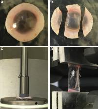
          Elastic properties (in-plane Young’s, E, and out-of-plane, G, shear modulus) of normal and ultraviolet CXL–treated human corneas were quantified using noncontact AμT-OCE. A nearly incompressible transverse isotropic model was used to reconstruct moduli from AμT-OCE data. Independently, cornea elastic moduli were also measured with destructive mechanical tests (tensile extensometry and shear rheometry).

          Main Outcome Measures

          Corneal elastic moduli (in-plane Young’s modulus, E, in-plane, μ, and out-of-plane, G, shear moduli) can be evaluated in both normal and CXL treated tissues, as well as monitored during the CXL procedure using noncontact AμT-OCE.

          Results

          Cross-linking induced a significant increase in both in-plane and out-of-plane elastic moduli in human cornea. The statistical mean in the paired study (presurgery and postsurgery, n = 7) of the in-plane Young’s modulus, E = 3 μ , increased from 19 MPa to 43 MPa, while the out-of-plane shear modulus, G, increased from 188 kPa to 673 kPa. Mechanical tests in a separate subgroup support CXL-induced cornea moduli changes and generally agree with noncontact AμT-OCE measurements.

          Conclusions

          The human cornea is a highly anisotropic material where in-plane mechanical properties are very different from those out-of-plane. Noncontact AμT-OCE can measure changes in the anisotropic elastic properties in human cornea as a result of ultraviolet CXL.

          Related collections

          Most cited references76

          • Record: found
          • Abstract: found
          • Article: not found

          Stress-strain measurements of human and porcine corneas after riboflavin-ultraviolet-A-induced cross-linking.

          To evaluate the biomechanical effect of combined riboflavin-ultraviolet A (UVA) treatment on porcine and human corneas. Department of Ophthalmology, Technical University of Dresden, Dresden, Germany. Corneal strips from 5 human enucleated eyes and 20 porcine cadaver corneas were treated with the photosensitizer riboflavin and irradiated with 2 double UVA diodes (370 nm, irradiance = 3 mW/cm2) for 30 minutes. After cross-linking, static stress-strain measurements of the treated and untreated corneas were performed using a microcomputer-controlled biomaterial tester with a prestress of 5 x 10(3) Pa. There was a significant increase in corneal rigidity after cross-linking, indicated by a rise in stress in treated porcine corneas (by 71.9%) and human corneas (by 328.9%) and in Young's modulus by the factor 1.8 in porcine corneas and 4.5 in human corneas. The mean central corneal thickness was 850 microm +/- 70 (SD) in porcine corneas and 550 +/- 40 microm in human corneas. Riboflavin-UVA-induced collagen cross-linking led to an increase in mechanical rigidity in porcine corneas and an even greater increase in human corneas. As collagen cross-linking is maximal in the anterior 300 microm of the cornea, the greater stiffening effect in human corneas can be explained by the relatively larger portion of the cornea being cross-linked in the overall thinner human cornea.
            Bookmark
            • Record: found
            • Abstract: found
            • Article: not found

            Safety of UVA-riboflavin cross-linking of the cornea.

            To study potential damage to ocular tissue during corneal collagen cross-linking (X-linking) by means of the riboflavin/UVA (370 nm) approach. Comparison of the currently used technique with officially accepted guidelines regarding direct UV damage and the damage created by the induced free radicals (photochemical damage). The currently used UVA radiant exposure of 5.4 mJ/cm and the corresponding irradiance of 3 mW/cm2 is below the known damage thresholds of UVA for the corneal endothelium, lens, and retina. Regarding the photochemical damage caused by the free radicals, the damage thresholds for keratocytes and endothelial cells are 0.45 and 0.35 mW/cm, respectively. In a 400-microm-thick cornea saturated with riboflavin, the irradiance at the endothelial level was 0.18 mW/cm, which is a factor of 2 smaller than the damage threshold. After corneal X-linking, the stroma is depopulated of keratocytes approximately 300 microm deep. Repopulation of this area takes up to 6 months. As long as the cornea treated has a minimum thickness of 400 microm (as recommended), the corneal endothelium will not experience damage, nor will deeper structures such as lens and retina. The light source should provide a homogenous irradiance, avoiding hot spots.
              Bookmark
              • Record: found
              • Abstract: found
              • Article: not found

              Riboflavin/ultraviolet-a-induced collagen crosslinking for the treatment of keratoconus.

              In animal eyes, a significant increase in corneal biomechanical stiffness has been found after collagen crosslinking by combined riboflavin/ultraviolet-A (UVA) treatment. The aim of the present study was to evaluate the clinical usefulness of riboflavin/UVA-induced collagen crosslinking for bringing the progression of keratoconus to a halt. Prospective, nonrandomized clinical pilot study. Twenty-three eyes of 22 patients with moderate or advanced progressive keratoconus (maximum K value, 48-72 diopters) were included. After central corneal abrasion, photosensitizing riboflavin drops were applied and the eyes exposed to UVA (370 nm, 3 mW/cm(2)) in a 1-cm distance for 30 minutes. Postoperative examinations were performed in 6-month intervals, including visual acuity testing, corneal topography, slit-lamp examination, measurement of endothelial cell density, and photographic documentation. The follow-up time was between 3 months and 4 years. In all treated eyes, the progression of keratoconus was at least stopped. In 16 eyes (70%) regression with a reduction of the maximal keratometry readings by 2.01 diopters and of the refractive error by 1.14 diopters was found. Corneal and lens transparency, endothelial cell density, and intraocular pressure remained unchanged. Visual acuity improved slightly in 15 eyes (65%). Collagen crosslinking may be a new way for stopping the progression of keratectasia in patients with keratoconus. The need for penetrating keratoplasty might then be significantly reduced in keratoconus. Given the simplicity and minimal costs of the treatment, it might also be well-suited for developing countries. Long-term results are necessary to evaluate the duration of the stiffening effect and to exclude long term side-effects.
                Bookmark

                Author and article information

                Contributors
                Journal
                Ophthalmol Sci
                Ophthalmol Sci
                Ophthalmology Science
                Elsevier
                2666-9145
                13 November 2022
                June 2023
                13 November 2022
                : 3
                : 2
                : 100257
                Affiliations
                [1 ]Department of Bioengineering, University of Washington, Seattle, Washington
                [2 ]School of Medicine, University of Washington, Seattle, Washington
                [3 ]Department of Ophthalmology, University of Washington, Seattle, Washington
                Author notes
                []Correspondence: Tueng T. Shen, PhD, MD, UW Medicine Eye Institute, Seattle, Washington 98104. ttshen@ 123456uw.edu
                Article
                S2666-9145(22)00146-4 100257
                10.1016/j.xops.2022.100257
                9852959
                36685713
                048e3589-a755-4bf2-99e0-b1cf9a1ee623
                © 2022 Published by Elsevier Inc. on behalf of American Academy of Ophthalmology.

                This is an open access article under the CC BY-NC-ND license (http://creativecommons.org/licenses/by-nc-nd/4.0/).

                History
                : 15 September 2022
                : 25 October 2022
                : 8 November 2022
                Categories
                Original Article

                cornea,cross-linking,elastic anisotropy,niti model,optical coherence elastography,aμt, acoustic micro-tapping,bss, balanced saline solution,cxl, cross-linking,iop, intraocular pressure,niti, nearly incompressible transverse isotropy,oce, optical coherence elastography,rf, riboflavin,uv, ultraviolet

                Comments

                Comment on this article