4
views
0
recommends
+1 Recommend
0 collections
    0
    shares
      • Record: found
      • Abstract: found
      • Article: found
      Is Open Access

      Loss of mural cell-derived laminin aggravates hemorrhagic brain injury

      research-article

      Read this article at

      Bookmark
          There is no author summary for this article yet. Authors can add summaries to their articles on ScienceOpen to make them more accessible to a non-specialist audience.

          Abstract

          Background

          Mural cells synthesize and deposit laminin to the basement membrane. To investigate the function of mural cell-derived laminin, we generated a mutant mouse line lacking mural cell-derived laminin (termed PKO). In a previous study, we showed that the PKO mice were grossly normal under homeostatic condition, but developed blood-brain barrier (BBB) breakdown with advanced age (> 8 months), suggesting that these mutants are intrinsically weak. Based on these findings, we hypothesized that PKO mice have exacerbated injuries in pathological conditions.

          Methods

          Using collagenase-induced intracerebral hemorrhage (ICH) as an injury model, we examined various stroke outcomes, including hematoma volume, neurological function, neuronal death, BBB integrity, paracellular/transcellular transport, inflammatory cell infiltration, and brain water content, in PKO mice and their wildtype littermates at young age (6–8 weeks). In addition, transmission electron microscopy (TEM) analysis and an in vitro ICH model were used to investigate the underlying molecular mechanisms.

          Results

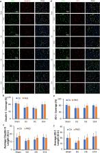
          Compared to age-matched wildtype littermates, PKO mice display aggravated stroke outcomes, including larger hematoma size, worse neurological function, increased neuronal cell death, enhanced BBB permeability, increased transcytosis, and elevated inflammatory cell infiltration. These mutants also exhibit high baseline brain water content independent of aquaporin-4 (AQP4). In addition, mural cell-derived laminin significantly reduced caveolin-1 without affecting tight junction proteins in the in vitro ICH model.

          Conclusions

          These results suggest that mural cell-derived laminin attenuates BBB damage in ICH via decreasing caveolin-1 and thus transcytosis, regulates brain water homeostasis, and plays a beneficial role in ICH.

          Related collections

          Most cited references69

          • Record: found
          • Abstract: not found
          • Article: not found

          Intracerebral haemorrhage: current approaches to acute management

            Bookmark
            • Record: found
            • Abstract: found
            • Article: not found

            Fluoro-Jade B: a high affinity fluorescent marker for the localization of neuronal degeneration.

            Fluoro-Jade B, like its predecessor Fluoro-Jade, is an anionic fluorescein derivative useful for the histological staining of neurons undergoing degeneration. However, Fluoro-Jade B has an even greater specific affinity for degenerating neurons. This notion is supported by the conspicuous staining of degenerating neuronal elements with minimal background staining. This improved signal-to-noise ratio means that fine neuronal processes including distal dendrites, axons and axon terminals can be more readily detected and documented. Although the staining time and dye concentration are reduced, the method is as rapid, simple and reliable as the original Fluoro-Jade technique. Like Fluoro-Jade, Fluoro-Jade B is compatible with a number of other labeling procedures including immunofluorescent and fluorescent Nissl techniques.
              Bookmark
              • Record: found
              • Abstract: found
              • Article: not found

              The neurovascular unit - concept review.

              The cerebral hyperaemia is one of the fundamental mechanisms for the central nervous system homeostasis. Due also to this mechanism, oxygen and nutrients are maintained in satisfactory levels, through vasodilation and vasoconstriction. The brain hyperaemia, or coupling, is accomplished by a group of cells, closely related to each other; called neurovascular unit (NVU). The neurovascular unit is composed by neurones, astrocytes, endothelial cells of blood-brain barrier (BBB), myocytes, pericytes and extracellular matrix components. These cells, through their intimate anatomical and chemical relationship, detect the needs of neuronal supply and trigger necessary responses (vasodilation or vasoconstriction) for such demands. Here, we review the concepts of NVU, the coupling mechanisms and research strategies. © 2014 Scandinavian Physiological Society. Published by John Wiley & Sons Ltd.
                Bookmark

                Author and article information

                Contributors
                yyao@uga.edu
                Journal
                J Neuroinflammation
                J Neuroinflammation
                Journal of Neuroinflammation
                BioMed Central (London )
                1742-2094
                6 April 2020
                6 April 2020
                2020
                : 17
                : 103
                Affiliations
                GRID grid.213876.9, ISNI 0000 0004 1936 738X, Department of Pharmaceutical and Biomedical Sciences, , University of Georgia, ; 240 W Green Street, Athens, GA 30602 USA
                Article
                1788
                10.1186/s12974-020-01788-3
                7133020
                32252790
                044ceaad-3ece-4b61-be24-907eba4c906d
                © The Author(s) 2020

                Open AccessThis article is licensed under a Creative Commons Attribution 4.0 International License, which permits use, sharing, adaptation, distribution and reproduction in any medium or format, as long as you give appropriate credit to the original author(s) and the source, provide a link to the Creative Commons licence, and indicate if changes were made. The images or other third party material in this article are included in the article's Creative Commons licence, unless indicated otherwise in a credit line to the material. If material is not included in the article's Creative Commons licence and your intended use is not permitted by statutory regulation or exceeds the permitted use, you will need to obtain permission directly from the copyright holder. To view a copy of this licence, visit http://creativecommons.org/licenses/by/4.0/. The Creative Commons Public Domain Dedication waiver ( http://creativecommons.org/publicdomain/zero/1.0/) applies to the data made available in this article, unless otherwise stated in a credit line to the data.

                History
                : 20 January 2020
                : 25 March 2020
                Funding
                Funded by: FundRef http://dx.doi.org/10.13039/100000050, National Heart, Lung, and Blood Institute;
                Award ID: R01HL146574
                Award Recipient :
                Funded by: FundRef http://dx.doi.org/10.13039/100000968, American Heart Association;
                Award ID: 16SDG29320001
                Award Recipient :
                Categories
                Research
                Custom metadata
                © The Author(s) 2020

                Neurosciences
                intracerebral hemorrhage,blood-brain barrier,mural cells,edema,laminin
                Neurosciences
                intracerebral hemorrhage, blood-brain barrier, mural cells, edema, laminin

                Comments

                Comment on this article