17
views
0
recommends
+1 Recommend
0 collections
    0
    shares
      • Record: found
      • Abstract: found
      • Article: found
      Is Open Access

      CEST MR fingerprinting (CEST‐MRF) for brain tumor quantification using EPI readout and deep learning reconstruction

      research-article

      Read this article at

      Bookmark
          There is no author summary for this article yet. Authors can add summaries to their articles on ScienceOpen to make them more accessible to a non-specialist audience.

          Abstract

          Purpose

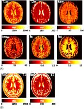
          To develop a clinical CEST MR fingerprinting (CEST‐MRF) method for brain tumor quantification using EPI acquisition and deep learning reconstruction.

          Methods

          A CEST‐MRF pulse sequence originally designed for animal imaging was modified to conform to hardware limits on clinical scanners while keeping scan time under 2 min. Quantitative MRF reconstruction was performed using a deep reconstruction network (DRONE) to yield the water relaxation and chemical exchange parameters. The feasibility of the six parameter DRONE reconstruction was tested in simulations using a digital brain phantom. A healthy subject was scanned with the CEST‐MRF sequence, conventional MRF and CEST sequences for comparison. Reproducibility was assessed via test–retest experiments and the concordance correlation coefficient calculated for white matter and gray matter. The clinical utility of CEST‐MRF was demonstrated on four patients with brain metastases in comparison to standard clinical imaging sequences. Tumors were segmented into edema, solid core, and necrotic core regions and the CEST‐MRF values compared to the contra‐lateral side.

          Results

          DRONE reconstruction of the digital phantom yielded a normalized RMS error of ≤7% for all parameters. The CEST‐MRF parameters were in good agreement with those from conventional MRF and CEST sequences and previous studies. The mean concordance correlation coefficient for all six parameters was 0.98 ± 0.01 in white matter and 0.98 ± 0.02 in gray matter. The CEST‐MRF values in nearly all tumor regions were significantly different ( P = 0.05) from each other and the contra‐lateral side.

          Conclusion

          Combination of EPI readout and deep learning reconstruction enabled fast, accurate and reproducible CEST‐MRF in brain tumors.

          Related collections

          Most cited references50

          • Record: found
          • Abstract: found
          • Article: not found

          PyTorch: An Imperative Style, High-Performance Deep Learning Library

          Deep learning frameworks have often focused on either usability or speed, but not both. PyTorch is a machine learning library that shows that these two goals are in fact compatible: it provides an imperative and Pythonic programming style that supports code as a model, makes debugging easy and is consistent with other popular scientific computing libraries, while remaining efficient and supporting hardware accelerators such as GPUs. In this paper, we detail the principles that drove the implementation of PyTorch and how they are reflected in its architecture. We emphasize that every aspect of PyTorch is a regular Python program under the full control of its user. We also explain how the careful and pragmatic implementation of the key components of its runtime enables them to work together to achieve compelling performance. We demonstrate the efficiency of individual subsystems, as well as the overall speed of PyTorch on several common benchmarks. 12 pages, 3 figures, NeurIPS 2019
            Bookmark
            • Record: found
            • Abstract: found
            • Article: not found

            A concordance correlation coefficient to evaluate reproducibility.

            L Lin (1989)
            A new reproducibility index is developed and studied. This index is the correlation between the two readings that fall on the 45 degree line through the origin. It is simple to use and possesses desirable properties. The statistical properties of this estimate can be satisfactorily evaluated using an inverse hyperbolic tangent transformation. A Monte Carlo experiment with 5,000 runs was performed to confirm the estimate's validity. An application using actual data is given.
              Bookmark
              • Record: found
              • Abstract: found
              • Article: not found

              Magnetic Resonance Fingerprinting

              Summary Magnetic Resonance (MR) is an exceptionally powerful and versatile measurement technique. The basic structure of an MR experiment has remained nearly constant for almost 50 years. Here we introduce a novel paradigm, Magnetic Resonance Fingerprinting (MRF) that permits the non-invasive quantification of multiple important properties of a material or tissue simultaneously through a new approach to data acquisition, post-processing and visualization. MRF provides a new mechanism to quantitatively detect and analyze complex changes that can represent physical alterations of a substance or early indicators of disease. MRF can also be used to specifically identify the presence of a target material or tissue, which will increase the sensitivity, specificity, and speed of an MR study, and potentially lead to new diagnostic testing methodologies. When paired with an appropriate pattern recognition algorithm, MRF inherently suppresses measurement errors and thus can improve accuracy compared to previous approaches.
                Bookmark

                Author and article information

                Contributors
                coheno1@mskcc.org
                Journal
                Magn Reson Med
                Magn Reson Med
                10.1002/(ISSN)1522-2594
                MRM
                Magnetic Resonance in Medicine
                John Wiley and Sons Inc. (Hoboken )
                0740-3194
                1522-2594
                21 September 2022
                January 2023
                : 89
                : 1 ( doiID: 10.1002/mrm.v89.1 )
                : 233-249
                Affiliations
                [ 1 ] Department of Medical Physics Memorial Sloan Kettering Cancer Center New York New York USA
                [ 2 ] Department of Radiation Oncology Memorial Sloan Kettering Cancer Center New York New York USA
                [ 3 ] Department of Radiology Memorial Sloan Kettering Cancer Center New York New York USA
                [ 4 ] Athinoula A. Martinos Center for Biomedical Imaging, Department of Radiology Massachusetts General Hospital and Harvard Medical School Charlestown Massachusetts USA
                [ 5 ] Department of Biomedical Engineering Tel Aviv University Tel Aviv Israel
                [ 6 ] Sagol School of Neuroscience Tel Aviv University Tel Aviv Israel
                Author notes
                [*] [* ] Correspondence

                Ouri Cohen, Memorial Sloan Kettering Cancer Center, 320 East 61 St, New York, NY 10025, USA.

                Email: coheno1@ 123456mskcc.org

                Author information
                https://orcid.org/0000-0003-3632-8094
                https://orcid.org/0000-0002-3566-569X
                https://orcid.org/0000-0002-3782-4930
                Article
                MRM29448
                10.1002/mrm.29448
                9617776
                36128888
                03ecb9df-d3c4-4845-90e4-9d4acfafa6ab
                © 2022 The Authors. Magnetic Resonance in Medicine published by Wiley Periodicals LLC on behalf of International Society for Magnetic Resonance in Medicine.

                This is an open access article under the terms of the http://creativecommons.org/licenses/by-nc/4.0/ License, which permits use, distribution and reproduction in any medium, provided the original work is properly cited and is not used for commercial purposes.

                History
                : 09 August 2022
                : 12 January 2022
                : 19 August 2022
                Page count
                Figures: 9, Tables: 1, Pages: 17, Words: 8453
                Funding
                Funded by: European Union , doi 10.13039/501100000780;
                Award ID: 836752
                Funded by: NIH/NCI
                Award ID: P30‐CA008748
                Award ID: R37CA262662‐01A1
                Categories
                Research Article
                Research Articles—Imaging Methodology
                Custom metadata
                2.0
                January 2023
                Converter:WILEY_ML3GV2_TO_JATSPMC version:6.2.3 mode:remove_FC converted:08.01.2023

                Radiology & Imaging
                chemical exchange rate,chemical exchange saturation transfer (cest),deep learning,drone,magnetic resonance fingerprinting (mrf),ph

                Comments

                Comment on this article