6
views
0
recommends
+1 Recommend
0 collections
    0
    shares
      • Record: found
      • Abstract: found
      • Article: found
      Is Open Access

      Multilevel Analysis of the Neovascularization and Integration Process of a Nonvascularized Rectus Fascia Transplantation

      research-article

      Read this article at

      Bookmark
          There is no author summary for this article yet. Authors can add summaries to their articles on ScienceOpen to make them more accessible to a non-specialist audience.

          Abstract

          Background.

          Failure to close the abdominal wall after intestinal transplantation (ITx) or multivisceral Tx remains a surgical challenge. An attractive method is the use of nonvascularized rectus fascia (NVRF) in which both layers of the donor abdominal rectus fascia are used as an inlay patch without vascular anastomosis. How this graft integrates over time remains unknown. The study aims to provide a multilevel analysis of the neovascularization and integration process of the NVRF.

          Methods.

          Three NVRF-Tx were performed after ITx. Clinical, radiological, histological, and immunological data were analyzed to get insights into the neovascularization and integration process of the NVRF. Moreover, cryogenic contrast-enhanced microfocus computed tomography (microCT) analysis was used for detailed reconstruction of the vasculature in and around the NVRF (3-dimensional histology).

          Results.

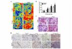
          Two men (31- and 51-y-old) and 1 woman (49-y-old) underwent 2 multivisceral Tx and 1 combined liver-ITx, respectively. A CT scan showed contrast enhancement around the fascia graft at 5 days post-Tx. At 6 weeks, newly formed blood vessels were visualized around the graft with Doppler ultrasound. Biopsies at 2 weeks post-Tx revealed inflammation around the NVRF and early fibrosis. At 6 months, classical 2-dimensional histological analysis of a biopsy confirmed integration of the fascia graft with strong fibrotic reaction without signs of rejection. A cryogenic contrast-enhanced microCT scan of the same biopsy revealed the presence of microvasculature, enveloping and penetrating the donor fascia.

          Conclusions.

          We showed clinical, histological, and microCT evidence of the neovascularization and integration process of the NVRF after Tx.

          Related collections

          Most cited references29

          • Record: found
          • Abstract: found
          • Article: not found

          Simultaneous three-dimensional visualization of mineralized and soft skeletal tissues by a novel microCT contrast agent with polyoxometalate structure.

          Biological tissues have a complex and heterogeneous 3D structure, which is only partially revealed by standard histomorphometry in 2D. We here present a novel chemical compound for contrast-enhanced microfocus computed tomography (CE-CT), a Hafnium-based Wells-Dawson polyoxometalate (Hf-POM), which allows simultaneous 3D visualization of mineralized and non-mineralized skeletal tissues, such as mineralized bone and bone marrow vasculature and adipocytes. We validated the novel contrast agent, which has a neutral pH in solution, by detailed comparison with (immuno)histology on murine long bones as blueprint, and showed that Hf-POM-based CE-CT can be used for virtual 3D histology. Furthermore, we quantified the 3D structure of the different skeletal tissues, as well as their spatial relation to each other, during aging and diet-induced obesity. We discovered, based on a single CE-CT dataset per sample, clear differences between the groups in bone structure, vascular network organization, characteristics of the adipose tissue and proximity of the different tissues to each other. These findings highlight the complementarity and added value of Hf-POM-based CE-CT compared to standard histomorphometry. As this novel technology provides a detailed 3D simultaneous representation of the structural organization of mineralized bone and bone marrow vasculature and adipose tissue, it will enable to improve insight in the interactions between these three tissues in several bone pathologies and to evaluate the in vivo performance of biomaterials for skeletal regeneration.
            Bookmark
            • Record: found
            • Abstract: found
            • Article: not found

            Transplantation of the abdominal wall.

            Closure of the abdomen in patients undergoing intestinal transplantation can be extremely difficult, if not impossible. We describe our initial experience with abdominal wall allotransplantation to facilitate abdominal closure. We undertook nine cadaveric abdominal wall composite allograft transplants in eight patients. The graft's blood supply was based on the inferior epigastric vessels left in continuity with the donor femoral and iliac vessels. Skin biopsies were undertaken randomly and when rejection was suspected. Vessel patency was monitored by doppler ultrasound. Six patients have survived, five of whom have intact, viable abdominal wall grafts. Two patients have had a clinically mild episode of acute rejection of the skin of the abdominal wall that resolved with corticosteroid therapy. No clinically apparent graft-versus-host disease has been noted. Transplantation of an abdominal wall composite allograft can facilitate reconstruction and closure of the abdominal compartment in intestinal transplant recipients with complex abdominal wall defects.
              Bookmark
              • Record: found
              • Abstract: found
              • Article: found
              Is Open Access

              Negative pressure wound therapy promotes vessel destabilization and maturation at various stages of wound healing and thus influences wound prognosis

              Negative pressure wound therapy (NPWT) has been observed to accelerate the wound healing process in humans through promoting angiogenesis. However, the potential biological effect and relevant molecular mechanisms, including microvessel destabilization, regression and endothelial cell proliferation in the early stage (1–3 days), and the neovascular stabilization and maturation in the later stage (7–15 days), have yet to be fully elucidated. The current study aimed to research the potential effect of NPWT on angiogenesis and vessel maturation, and investigate relevant association between mature microvessels and wound prognosis, as well as the regulatory mechanisms in human wound healing. Patients in the present study (n=48) were treated with NPWT or a petrolatum gauze, and relevant growth factors and vessel changes were detected using various experimental methods. NPWT increased the expression levels of angiogenin-2 (Ang-2), and decreased the expression levels of Ang-1 and ratios of Ang-1/Ang-2 in the initial stages of wound healing. However, in the latter stages of wound healing, NPWT increased the expression levels of Ang-1 and ratios of Ang-1/Ang-2, as well as the phosphorylation level of tyrosine kinase receptor-2. Consequently, microvessel pericyte coverage was gradually elevated, and the basement membrane was gradually supplied with new blood at the later stage of wound healing. In conclusion, NPWT may preferentially stimulate microvessel destabilization and regression in the early stage of wound healing, and as a consequence, increase angiogenesis. Subsequently, in the later stage of wound healing, NPWT may preferentially promote microvessel stabilization, thereby promoting microvessel maturation in human wounds through the angiogenin/tyrosine kinase receptor-2 signaling pathway. The results of the present study results demonstrated that NPWT was able to accelerate wound healing speed, and thus influence wound prognosis, as a result of an abundance of mature microvessels in human wounds.
                Bookmark

                Author and article information

                Contributors
                Journal
                Transplant Direct
                Transplant Direct
                TXD
                Transplantation Direct
                Lippincott Williams & Wilkins (Hagerstown, MD )
                2373-8731
                15 May 2024
                June 2024
                : 10
                : 6
                : e1624
                Affiliations
                [1 ] Leuven Intestinal Failure and Transplantation (LIFT) Center, University Hospitals Leuven, Leuven, Belgium.
                [2 ] Department of Abdominal Transplant Surgery, University Hospitals Leuven, Leuven, Belgium.
                [3 ] Department of Chronic Diseases and Metabolism, Laboratory of Respiratory Diseases and Thoracic Surgery (BREATHE), KU Leuven, Leuven, Belgium.
                [4 ] Department of Materials Engineering, KU Leuven, Leuven, Belgium.
                [5 ] Biomechanics Lab, Institute of Mechanics, Materials and Civil Engineering, UCLouvain, Louvain-la-Neuve, Belgium.
                [6 ] Pole of Morphology, Institute of Experimental and Clinical Research, UCLouvain, Brussels, Belgium.
                [7 ] Department of Pathology, University Hospitals Leuven, Leuven, Belgium.
                [8 ] Unit of Translational Cell- and Tissue Research, Department of Imaging and Pathology, KU Leuven, Leuven, Belgium.
                [9 ] Department of Abdominal Surgery, University Hospitals Leuven, Leuven, Belgium.
                [10 ] Department of Development and Regeneration, Unit of Urogenital, Abdominal and Plastic Surgery, KU Leuven, Leuven, Belgium.
                [11 ] Prometheus Division of Skeletal Tissue Engineering, KU Leuven, Leuven, Belgium.
                [12 ] Department of Microbiology, Immunology and Transplantation, KU Leuven, Leuven, Belgium.
                [13 ] Department of Radiology, University Hospitals Leuven, Leuven, Belgium.
                [14 ] Translational MRI Unit, Department of Imaging and Pathology, KU Leuven, Leuven, Belgium.
                [15 ] Tissue Engineering Lab, Department of Development and Regeneration, KU Leuven, KULAK campus Kortrijk, Kortrijk, Belgium.
                [16 ] Histocompatibility and Immunogenetics Laboratory, Belgian Red Cross-Flanders, Mechelen, Belgium.
                [17 ] Department of Abdominal Surgery, Sint-Franciscusziekenhuis, Heusden-Zolder, Belgium.
                [18 ] Nuffield Department of Surgical Sciences, University of Oxford, Oxford, United Kingdom.
                [19 ] Department of Gastroenterology and Hepatology, University Hospitals Leuven, Leuven, Belgium.
                [20 ] Department of Chronic Diseases and Metabolism, Translational Research Center for Gastrointestinal Disorders (TARGID), KU Leuven, Leuven, Belgium.
                [21 ] Department of Thoracic Surgery, University Hospitals Leuven, Leuven, Belgium.
                Author notes
                Correspondence: Laurens J. Ceulemans, MD, PhD, Department of Thoracic Surgery and Lung Transplantation, University Hospitals Leuven/KU Leuven, Herestraat 49, 3000 Leuven, Belgium. ( laurens.ceulemans@ 123456uzleuven.be ); LinkedIn: www.linkedin.com/in/laurens-ceulemans-1190a7a1; Twitter: @CeulemansLJ.
                Author information
                https://orcid.org/0000-0002-0646-2615
                https://orcid.org/0000-0002-1488-5079
                https://orcid.org/0000-0001-8494-7725
                https://orcid.org/0000-0003-4862-478X
                https://orcid.org/0000-0002-1750-8324
                https://orcid.org/0000-0002-2839-8896
                https://orcid.org/0000-0002-0800-3279
                https://orcid.org/0000-0002-5896-6823
                https://orcid.org/0000-0002-0360-5385
                https://orcid.org/0000-0002-2653-8656
                https://orcid.org/0000-0003-0176-454X
                https://orcid.org/0000-0002-0506-1609
                https://orcid.org/0000-0003-4921-466X
                https://orcid.org/0000-0001-8728-0903
                https://orcid.org/0000-0002-8147-8801
                https://orcid.org/0000-0002-4261-7100
                Article
                TXD-2024-0016 00007
                10.1097/TXD.0000000000001624
                11098214
                38757048
                03d9de83-1a5e-4eb3-a75c-d846cccbbc3c
                Copyright © 2024 The Author(s). Transplantation Direct. Published by Wolters Kluwer Health, Inc.

                This is an open-access article distributed under the terms of the Creative Commons Attribution-Non Commercial-No Derivatives License 4.0 (CCBY-NC-ND), where it is permissible to download and share the work provided it is properly cited. The work cannot be changed in any way or used commercially without permission from the journal.

                History
                : 22 January 2024
                : 05 February 2024
                Categories
                Intestinal Transplantation
                Custom metadata
                TRUE
                T

                Comments

                Comment on this article