67
views
0
recommends
+1 Recommend
0 collections
    0
    shares
      • Record: found
      • Abstract: found
      • Article: found
      Is Open Access

      High expression of the putative cancer stem cell marker, DCLK1, in rectal neuroendocrine tumors

      research-article

      Read this article at

      Bookmark
          There is no author summary for this article yet. Authors can add summaries to their articles on ScienceOpen to make them more accessible to a non-specialist audience.

          Abstract

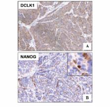
          Doublecortin-like kinase 1 (DCLK1), a microtubule-associated protein, is known to regulate neuronal differentiation, migration and neurogenesis. Recent evidence suggests that the protein is a putative marker for intestinal and pancreatic stem cells, including their cancer stem cell counterparts. The present study conducted immunohistochemical analyses for DCLK1 and the stemness marker, NANOG, in human intestinal neuroendocrine tumors (NETs), as their expression had not been previously investigated in these tumors. Eighteen patients with endoscopically resected rectal NETs were enrolled in the study. The mean age of the patients was 51 years old. The mean diameter of the resected tumors was 5.2 mm, and a histological diagnosis of NET grade G1 was formed for all tumors. Immunohistochemical analysis was performed not only for DCLK1, but also for the known NET markers, synaptophysin, chromogranin A and cluster of differentiation (CD)56. The intensity and distribution of staining were scored on a scale of 0–3 and 0–2, respectively. The sum of the scores was calculated for each specimen. Co-expression of DCLK1 and NANOG was also examined. The mean scores for DCLK1 and synaptophysin were significantly higher than those for chromogranin A (P<0.0001) and CD56 (P<0.01). There were no significant differences in the scores between DCLK1 and synaptophysin or between chromogranin A and CD56. Notably, NANOG was expressed in high quantities in all the tumor tissues studied, showing clear co-expression with DCLK1. In conclusion, DCLK1 may be a novel marker for rectal NET, potentially indicating the presence of the stemness gene product, NANOG.

          Related collections

          Most cited references26

          • Record: found
          • Abstract: found
          • Article: not found

          Dclk1 distinguishes between tumor and normal stem cells in the intestine.

          There is great interest in tumor stem cells (TSCs) as potential therapeutic targets; however, cancer therapies targeting TSCs are limited. A drawback is that TSC markers are often shared by normal stem cells (NSCs); thus, therapies that target these markers may cause severe injury to normal tissues. To identify a potential TSC-specific marker, we focused on doublecortin-like kinase 1 (Dclk1). Dclk1 was reported as a candidate NSC marker in the gut, but recent reports have implicated it as a marker of differentiated cells (for example, Tuft cells). Using lineage-tracing experiments, we show here that Dclk1 does not mark NSCs in the intestine but instead marks TSCs that continuously produce tumor progeny in the polyps of Apc(Min/+) mice. Specific ablation of Dclk1-positive TSCs resulted in a marked regression of polyps without apparent damage to the normal intestine. Our data suggest the potential for developing a therapy for colorectal cancer based on targeting Dclk1-positive TSCs.
            Bookmark
            • Record: found
            • Abstract: found
            • Article: not found

            Long-lived intestinal tuft cells serve as colon cancer-initiating cells.

            Doublecortin-like kinase 1 protein (DCLK1) is a gastrointestinal tuft cell marker that has been proposed to identify quiescent and tumor growth-sustaining stem cells. DCLK1⁺ tuft cells are increased in inflammation-induced carcinogenesis; however, the role of these cells within the gastrointestinal epithelium and their potential as cancer-initiating cells are poorly understood. Here, using a BAC-CreERT-dependent genetic lineage-tracing strategy, we determined that a subpopulation of DCLK1⁺ cells is extremely long lived and possesses rare stem cell abilities. Moreover, genetic ablation of Dclk1 revealed that DCLK1⁺ tuft cells contribute to recovery following intestinal and colonic injury. Surprisingly, conditional knockdown of the Wnt regulator APC in DCLK1⁺ cells was not sufficient to drive colonic carcinogenesis under normal conditions; however, dextran sodium sulfate-induced (DSS-induced) colitis promoted the development of poorly differentiated colonic adenocarcinoma in mice lacking APC in DCLK1⁺ cells. Importantly, colonic tumor formation occurred even when colitis onset was delayed for up to 3 months after induced APC loss in DCLK1⁺ cells. Thus, our data define an intestinal DCLK1⁺ tuft cell population that is long lived, quiescent, and important for intestinal homeostasis and regeneration. Long-lived DCLK1⁺ cells maintain quiescence even following oncogenic mutation, but are activated by tissue injury and can serve to initiate colon cancer.
              Bookmark
              • Record: found
              • Abstract: found
              • Article: not found

              DCLK1 marks a morphologically distinct subpopulation of cells with stem cell properties in preinvasive pancreatic cancer.

              As in other tumor types, progression of pancreatic cancer may require a functionally unique population of cancer stem cells. Although such cells have been identified in many invasive cancers, it is not clear whether they emerge during early or late stages of tumorigenesis. Using mouse models and human pancreatic cancer cell lines, we investigated whether preinvasive pancreatic neoplasia contains a subpopulation of cells with distinct morphologies and cancer stem cell-like properties. Pancreatic tissue samples were collected from the KC(Pdx1), KPC(Pdx1), and KC(iMist1) mouse models of pancreatic intraepithelial neoplasia (PanIN) and analyzed by confocal and electron microscopy, lineage tracing, and fluorescence-activated cell sorting. Subpopulations of human pancreatic ductal adenocarcinoma (PDAC) cells were similarly analyzed and also used in complementary DNA microarray analyses. The microtubule regulator DCLK1 marked a morphologically distinct and functionally unique population of pancreatic cancer-initiating cells. These cells displayed morphological and molecular features of gastrointestinal tuft cells. Cells that expressed DCLK1 also expressed high levels of ATAT1, HES1, HEY1, IGF1R, and ABL1, and manipulation of these pathways in PDAC cell lines inhibited their clonogenic potential. Pharmacological inhibition of γ-secretase activity reduced the abundance of these cells in murine PanIN in a manner that correlated with inhibition of PanIN progression. Human PDAC cells and pancreatic neoplasms in mice contain morphologically and functionally distinct subpopulations that have cancer stem cell-like properties. These populations can be identified at the earliest stages of pancreatic tumorigenesis and provide new cellular and molecular targets for pancreatic cancer treatment and/or chemoprevention. Copyright © 2014 AGA Institute. Published by Elsevier Inc. All rights reserved.
                Bookmark

                Author and article information

                Journal
                Oncol Lett
                Oncol Lett
                OL
                Oncology Letters
                D.A. Spandidos
                1792-1074
                1792-1082
                October 2015
                20 July 2015
                20 July 2015
                : 10
                : 4
                : 2015-2020
                Affiliations
                [1 ]Division of Gastroenterology, Department of Medicine, Kurume University School of Medicine, Kurume, Japan
                [2 ]Research Center for Innovative Cancer Therapy, Kurume University, Kurume, Japan
                [3 ]Department of Pathology, Kurume University School of Medicine, Kurume, Japan
                [4 ]Department of Diagnostic Pathology, Kurume University Hospital, Kurume, Japan
                Author notes
                Correspondence to: Dr Hironori Koga, Division of Gastroenterology, Department of Medicine, Kurume University School of Medicine, 67 Asahi-Machi, Kurume 830-0011, Japan, E-mail: hirokoga@ 123456med.kurume-u.ac.jp
                Article
                OL-0-0-3513
                10.3892/ol.2015.3513
                4579808
                03d937be-4f44-4b84-94bd-f9601eabb59f
                Copyright: © Ikezono et al. This is an open access article distributed under the terms of a Creative Commons Attribution License.

                This is an open-access article licensed under a Creative Commons Attribution-NonCommercial 4.0 Unported License. The article may be redistributed, reproduced, and reused for non-commercial purposes, provided the original source is properly cited.

                History
                : 31 August 2014
                : 15 May 2015
                Categories
                Articles

                Oncology & Radiotherapy
                carcinoids,neuroendocrine tumor,cancer stem cell,nanog
                Oncology & Radiotherapy
                carcinoids, neuroendocrine tumor, cancer stem cell, nanog

                Comments

                Comment on this article