10
views
0
recommends
+1 Recommend
0 collections
    0
    shares
      • Record: found
      • Abstract: found
      • Article: found
      Is Open Access

      EARL compliance and imaging optimisation on the Biograph Vision Quadra PET/CT using phantom and clinical data

      research-article

      Read this article at

      Bookmark
          There is no author summary for this article yet. Authors can add summaries to their articles on ScienceOpen to make them more accessible to a non-specialist audience.

          Abstract

          Purpose

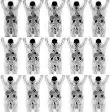
          Current European Association of Nuclear Medicine (EANM) Research Ltd. (EARL) guidelines for the standardisation of PET imaging developed for conventional systems have not yet been adjusted for long axial field-of-view (LAFOV) systems. In order to use the LAFOV Siemens Biograph Vision Quadra PET/CT (Siemens Healthineers, Knoxville, TN, USA) in multicentre research and harmonised clinical use, compliance to EARL specifications for 18F-FDG tumour imaging was explored in the current study. Additional tests at various locations throughout the LAFOV and the use of shorter scan durations were included. Furthermore, clinical data were collected to further explore and validate the effects of reducing scan duration on semi-quantitative PET image biomarker accuracy and precision when using EARL-compliant reconstruction settings.

          Methods

          EARL compliance phantom measurements were performed using the NEMA image quality phantom both in the centre and at various locations throughout the LAFOV. PET data (maximum ring difference (MRD) = 85) were reconstructed using various reconstruction parameters and reprocessed to obtain images at shorter scan durations. Maximum, mean and peak activity concentration recovery coefficients (RC) were obtained for each sphere and compared to EARL standards specifications.

          Additionally, PET data (MRD = 85) of 10 oncological patients were acquired and reconstructed using various reconstruction settings and reprocessed from 10 min listmode acquisition into shorter scan durations. Per dataset, SUVs were derived from tumour lesions and healthy tissues. ANOVA repeated measures were performed to explore differences in lesion SUV max and SUV peak. Wilcoxon signed-rank tests were performed to evaluate differences in background SUV peak and SUV mean between scan durations. The coefficient of variation (COV) was calculated to characterise noise.

          Results

          Phantom measurements showed EARL compliance for all positions throughout the LAFOV for all scan durations. Regarding patient data, EARL-compliant images showed no clinically meaningful significant differences in lesion SUV max and SUV peak or background SUV mean and SUV peak between scan durations. Here, COV only varied slightly.

          Conclusion

          Images obtained using the Vision Quadra PET/CT comply with EARL specifications. Scan duration and/or activity administration can be reduced up to a factor tenfold without the interference of increased noise.

          Supplementary Information

          The online version contains supplementary material available at 10.1007/s00259-022-05919-1.

          Related collections

          Most cited references21

          • Record: found
          • Abstract: found
          • Article: not found

          From RECIST to PERCIST: Evolving Considerations for PET response criteria in solid tumors.

          The purpose of this article is to review the status and limitations of anatomic tumor response metrics including the World Health Organization (WHO) criteria, the Response Evaluation Criteria in Solid Tumors (RECIST), and RECIST 1.1. This article also reviews qualitative and quantitative approaches to metabolic tumor response assessment with (18)F-FDG PET and proposes a draft framework for PET Response Criteria in Solid Tumors (PERCIST), version 1.0. PubMed searches, including searches for the terms RECIST, positron, WHO, FDG, cancer (including specific types), treatment response, region of interest, and derivative references, were performed. Abstracts and articles judged most relevant to the goals of this report were reviewed with emphasis on limitations and strengths of the anatomic and PET approaches to treatment response assessment. On the basis of these data and the authors' experience, draft criteria were formulated for PET tumor response to treatment. Approximately 3,000 potentially relevant references were screened. Anatomic imaging alone using standard WHO, RECIST, and RECIST 1.1 criteria is widely applied but still has limitations in response assessments. For example, despite effective treatment, changes in tumor size can be minimal in tumors such as lymphomas, sarcoma, hepatomas, mesothelioma, and gastrointestinal stromal tumor. CT tumor density, contrast enhancement, or MRI characteristics appear more informative than size but are not yet routinely applied. RECIST criteria may show progression of tumor more slowly than WHO criteria. RECIST 1.1 criteria (assessing a maximum of 5 tumor foci, vs. 10 in RECIST) result in a higher complete response rate than the original RECIST criteria, at least in lymph nodes. Variability appears greater in assessing progression than in assessing response. Qualitative and quantitative approaches to (18)F-FDG PET response assessment have been applied and require a consistent PET methodology to allow quantitative assessments. Statistically significant changes in tumor standardized uptake value (SUV) occur in careful test-retest studies of high-SUV tumors, with a change of 20% in SUV of a region 1 cm or larger in diameter; however, medically relevant beneficial changes are often associated with a 30% or greater decline. The more extensive the therapy, the greater the decline in SUV with most effective treatments. Important components of the proposed PERCIST criteria include assessing normal reference tissue values in a 3-cm-diameter region of interest in the liver, using a consistent PET protocol, using a fixed small region of interest about 1 cm(3) in volume (1.2-cm diameter) in the most active region of metabolically active tumors to minimize statistical variability, assessing tumor size, treating SUV lean measurements in the 1 (up to 5 optional) most metabolically active tumor focus as a continuous variable, requiring a 30% decline in SUV for "response," and deferring to RECIST 1.1 in cases that do not have (18)F-FDG avidity or are technically unsuitable. Criteria to define progression of tumor-absent new lesions are uncertain but are proposed. Anatomic imaging alone using standard WHO, RECIST, and RECIST 1.1 criteria have limitations, particularly in assessing the activity of newer cancer therapies that stabilize disease, whereas (18)F-FDG PET appears particularly valuable in such cases. The proposed PERCIST 1.0 criteria should serve as a starting point for use in clinical trials and in structured quantitative clinical reporting. Undoubtedly, subsequent revisions and enhancements will be required as validation studies are undertaken in varying diseases and treatments.
            Bookmark
            • Record: found
            • Abstract: found
            • Article: not found

            FDG PET/CT: EANM procedure guidelines for tumour imaging: version 2.0

            The purpose of these guidelines is to assist physicians in recommending, performing, interpreting and reporting the results of FDG PET/CT for oncological imaging of adult patients. PET is a quantitative imaging technique and therefore requires a common quality control (QC)/quality assurance (QA) procedure to maintain the accuracy and precision of quantitation. Repeatability and reproducibility are two essential requirements for any quantitative measurement and/or imaging biomarker. Repeatability relates to the uncertainty in obtaining the same result in the same patient when he or she is examined more than once on the same system. However, imaging biomarkers should also have adequate reproducibility, i.e. the ability to yield the same result in the same patient when that patient is examined on different systems and at different imaging sites. Adequate repeatability and reproducibility are essential for the clinical management of patients and the use of FDG PET/CT within multicentre trials. A common standardised imaging procedure will help promote the appropriate use of FDG PET/CT imaging and increase the value of publications and, therefore, their contribution to evidence-based medicine. Moreover, consistency in numerical values between platforms and institutes that acquire the data will potentially enhance the role of semiquantitative and quantitative image interpretation. Precision and accuracy are additionally important as FDG PET/CT is used to evaluate tumour response as well as for diagnosis, prognosis and staging. Therefore both the previous and these new guidelines specifically aim to achieve standardised uptake value harmonisation in multicentre settings.
              Bookmark
              • Record: found
              • Abstract: found
              • Article: not found

              FDG PET and PET/CT: EANM procedure guidelines for tumour PET imaging: version 1.0

              The aim of this guideline is to provide a minimum standard for the acquisition and interpretation of PET and PET/CT scans with [18F]-fluorodeoxyglucose (FDG). This guideline will therefore address general information about [18F]-fluorodeoxyglucose (FDG) positron emission tomography-computed tomography (PET/CT) and is provided to help the physician and physicist to assist to carrying out, interpret, and document quantitative FDG PET/CT examinations, but will concentrate on the optimisation of diagnostic quality and quantitative information.
                Bookmark

                Author and article information

                Contributors
                j.van.sluis@umcg.nl
                Journal
                Eur J Nucl Med Mol Imaging
                Eur J Nucl Med Mol Imaging
                European Journal of Nuclear Medicine and Molecular Imaging
                Springer Berlin Heidelberg (Berlin/Heidelberg )
                1619-7070
                1619-7089
                25 July 2022
                25 July 2022
                2022
                : 49
                : 13
                : 4652-4660
                Affiliations
                [1 ]GRID grid.4494.d, ISNI 0000 0000 9558 4598, Department of Nuclear Medicine and Molecular Imaging, , University of Groningen, University Medical Center Groningen, ; Groningen, The Netherlands
                [2 ]GRID grid.509540.d, ISNI 0000 0004 6880 3010, Department of Radiology and Nuclear Medicine, , Amsterdam University Medical Centers, ; location VUMC, Amsterdam, The Netherlands
                Author information
                http://orcid.org/0000-0003-1908-4518
                Article
                5919
                10.1007/s00259-022-05919-1
                9606094
                35876867
                03a19e5d-48c1-4d0d-8d46-254e6a7bab8e
                © The Author(s) 2022

                Open AccessThis article is licensed under a Creative Commons Attribution 4.0 International License, which permits use, sharing, adaptation, distribution and reproduction in any medium or format, as long as you give appropriate credit to the original author(s) and the source, provide a link to the Creative Commons licence, and indicate if changes were made. The images or other third party material in this article are included in the article's Creative Commons licence, unless indicated otherwise in a credit line to the material. If material is not included in the article's Creative Commons licence and your intended use is not permitted by statutory regulation or exceeds the permitted use, you will need to obtain permission directly from the copyright holder. To view a copy of this licence, visit http://creativecommons.org/licenses/by/4.0/.

                History
                : 6 April 2022
                : 16 July 2022
                Categories
                Original Article
                Custom metadata
                © Springer-Verlag GmbH Germany, part of Springer Nature 2022

                Radiology & Imaging
                imaging optimization,earl,scan duration,sipm,lafov pet/ct
                Radiology & Imaging
                imaging optimization, earl, scan duration, sipm, lafov pet/ct

                Comments

                Comment on this article