18
views
0
recommends
+1 Recommend
0 collections
    0
    shares
      • Record: found
      • Abstract: found
      • Article: found
      Is Open Access

      Aortic Valve Stenosis Alters Expression of Regional Aortic Wall Shear Stress: New Insights From a 4‐Dimensional Flow Magnetic Resonance Imaging Study of 571 Subjects

      research-article

      Read this article at

      Bookmark
          There is no author summary for this article yet. Authors can add summaries to their articles on ScienceOpen to make them more accessible to a non-specialist audience.

          Abstract

          Background

          Wall shear stress ( WSS) is a stimulus for vessel wall remodeling. Differences in ascending aorta ( AAo) hemodynamics have been reported between bicuspid aortic valve ( BAV) and tricuspid aortic valve patients with aortic dilatation, but the confounding impact of aortic valve stenosis ( AS) is unknown.

          Methods and Results

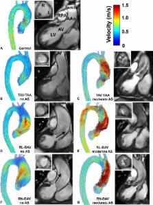
          Five hundred seventy‐one subjects underwent 4‐dimensional flow magnetic resonance imaging in the thoracic aorta (210 right‐left BAV cusp fusions, 60 right‐noncoronary BAV cusp fusions, 245 tricuspid aortic valve patients with aortic dilatation, and 56 healthy controls). There were 166 of 515 (32%) patients with AS. WSS atlases were created to quantify group‐specific WSS patterns in the AAo as a function of AS severity. In BAV patients without AS, the different cusp fusion phenotypes resulted in distinct differences in eccentric WSS elevation: right‐left BAV patients exhibited increased WSS by 9% to 34% ( P<0.001) at the aortic root and along the entire outer curvature of the AAo whereas right‐noncoronary BAV patients showed 30% WSS increase ( P<0.001) at the distal portion of the AAo. WSS in tricuspid aortic valve patients with aortic dilatation patients with no AS was significantly reduced by 21% to 33% ( P<0.01) in 4 of 6 AAo regions. In all patient groups, mild, moderate, and severe AS resulted in a marked increase in regional WSS ( P<0.001). Moderate‐to‐severe AS further increased WSS magnitude and variability in the AAo. Differences between valve phenotypes were no longer apparent.

          Conclusions

          AS significantly alters aortic hemodynamics and WSS independent of aortic valve phenotype and over‐rides previously described flow patterns associated with BAV and tricuspid aortic valve with aortic dilatation. Severity of AS must be considered when investigating valve‐mediated aortopathy.

          Related collections

          Most cited references33

          • Record: found
          • Abstract: not found
          • Article: not found

          2010 ACCF/AHA/AATS/ACR/ASA/SCA/SCAI/SIR/STS/SVM guidelines for the diagnosis and management of patients with Thoracic Aortic Disease: a report of the American College of Cardiology Foundation/American Heart Association Task Force on Practice Guidelines, American Association for Thoracic Surgery, American College of Radiology, American Stroke Association, Society of Cardiovascular Anesthesiologists, Society for Cardiovascular Angiography and Interventions, Society of Interventional Radiology, Society of Thoracic Surgeons, and Society for Vascular Medicine.

            Bookmark
            • Record: found
            • Abstract: found
            • Article: not found

            A classification system for the bicuspid aortic valve from 304 surgical specimens.

            In general, classification of a disease has proven to be advantageous for disease management. This may also be valid for the bicuspid aortic valve, because the term "bicuspid aortic valve" stands for a common congenital aortic valve malformation with heterogeneous morphologic phenotypes and function resulting in different treatment strategies. We attempted to establish a classification system based on a 5-year data collection of surgical specimens. Between 1999 and 2003 a precise description of valve pathology was obtained from operative reports of 304 patients with a diseased bicuspid aortic valve. Several different characteristics of bicuspid aortic valves were tested to generate a pithy and easily applicable classification system. Three characteristics for a systematic classification were found appropriate: (1) number of raphes, (2) spatial position of cusps or raphes, and (3) functional status of the valve. The first characteristic was found to be the most significant and therefore termed "type." Three major types were identified: type 0 (no raphe), type 1 (one raphe), and type 2 (two raphes), followed by two supplementary characteristics, spatial position and function. These characteristics served to classify and codify the bicuspid aortic valves into three categories. Most frequently, a bicuspid aortic valve with one raphe was identified (type 1, n = 269). This raphe was positioned between the left (L) and right (R) coronary sinuses in 216 (type 1, L/R) with a hemodynamic predominant stenosis (S) in 119 (type 1, L/R, S). Only 21 patients had a "purely" bicuspid aortic valve with no raphe (type 0). A classification system for the bicuspid aortic valve with one major category ("type") and two supplementary categories is presented. This classification, even if used in the major category (type) alone, might be advantageous to better define bicuspid aortic valve disease, facilitate scientific communication, and improve treatment.
              Bookmark
              • Record: found
              • Abstract: found
              • Article: not found

              Incidence of aortic complications in patients with bicuspid aortic valves.

              Bicuspid aortic valve (BAV), the most common congenital heart defect, has been thought to cause frequent and severe aortic complications; however, long-term, population-based data are lacking. To determine the incidence of aortic complications in patients with BAV in a community cohort and in the general population. In this retrospective cohort study, we conducted comprehensive assessment of aortic complications of patients with BAV living in a population-based setting in Olmsted County, Minnesota. We analyzed long-term follow-up of a cohort of all Olmsted County residents diagnosed with definite BAV by echocardiography from 1980 to 1999 and searched for aortic complications of patients whose bicuspid valves had gone undiagnosed. The last year of follow-up was 2008-2009. Thoracic aortic dissection, ascending aortic aneurysm, and aortic surgery. The cohort included 416 consecutive patients with definite BAV diagnosed by echocardiography, mean (SD) follow-up of 16 (7) years (6530 patient-years). Aortic dissection occurred in 2 of 416 patients; incidence of 3.1 (95% CI, 0.5-9.5) cases per 10,000 patient-years, age-adjusted relative-risk 8.4 (95% CI, 2.1-33.5; P = .003) compared with the county's general population. Aortic dissection incidences for patients 50 years or older at baseline and bearers of aortic aneurysms at baseline were 17.4 (95% CI, 2.9-53.6) and 44.9 (95% CI, 7.5-138.5) cases per 10,000 patient-years, respectively. Comprehensive search for aortic dissections in undiagnosed bicuspid valves revealed 2 additional patients, allowing estimation of aortic dissection incidence in bicuspid valve patients irrespective of diagnosis status (1.5; 95% CI, 0.4-3.8 cases per 10,000 patient-years), which was similar to the diagnosed cohort. Of 384 patients without baseline aneurysms, 49 developed aneurysms at follow-up, incidence of 84.9 (95% CI, 63.3-110.9) cases per 10,000 patient-years and an age-adjusted relative risk 86.2 (95% CI, 65.1-114; P <.001 compared with the general population). The 25-year rate of aortic surgery was 25% (95% CI, 17.2%-32.8%). In the population of patients with BAV, the incidence of aortic dissection over a mean of 16 years of follow-up was low but significantly higher than in the general population.
                Bookmark

                Author and article information

                Contributors
                alex.barker@northwestern.edu
                Journal
                J Am Heart Assoc
                J Am Heart Assoc
                10.1002/(ISSN)2047-9980
                JAH3
                ahaoa
                Journal of the American Heart Association: Cardiovascular and Cerebrovascular Disease
                John Wiley and Sons Inc. (Hoboken )
                2047-9980
                13 September 2017
                September 2017
                : 6
                : 9 ( doiID: 10.1002/jah3.2017.6.issue-9 )
                : e005959
                Affiliations
                [ 1 ] Department of Radiology Academic Medical Center Amsterdam The Netherlands
                [ 2 ] Department of Radiology Northwestern University Feinberg School of Medicine Chicago IL
                [ 3 ] Department of Medicine‐Cardiology Northwestern University Feinberg School of Medicine Chicago IL
                [ 4 ] Division of Surgery‐Cardiac Surgery Northwestern University Feinberg School of Medicine Chicago IL
                [ 5 ] Department of Biomedical Engineering Northwestern University Chicago IL
                [ 6 ] Department of Medical Imaging Ann & Robert H Lurie Children's Hospital of Chicago IL
                [ 7 ] Department of Cardiac Sciences University of Calgary Canada
                Author notes
                [*] [* ] Correspondence to: Alex J. Barker, PhD, Department of Radiology, Northwestern University Feinberg School of Medicine, 737 N Michigan Ave, Suite 1600, Chicago, IL 60611. E‐mail: alex.barker@ 123456northwestern.edu
                Article
                JAH32502
                10.1161/JAHA.117.005959
                5634265
                28903936
                024944d5-01d6-4e6a-923a-172e235e6b45
                © 2017 The Authors. Published on behalf of the American Heart Association, Inc., by Wiley.

                This is an open access article under the terms of the Creative Commons Attribution‐NonCommercial License, which permits use, distribution and reproduction in any medium, provided the original work is properly cited and is not used for commercial purposes.

                History
                : 03 April 2017
                : 12 July 2017
                Page count
                Figures: 4, Tables: 3, Pages: 13, Words: 9086
                Funding
                Funded by: National Heart, Lung, and Blood Institute
                Funded by: NIH
                Award ID: K25HL119608
                Award ID: R01HL115828,
                Award ID: R01HL133504
                Funded by: Martha and Richard Melman Family Bicuspid Aortic Valve Program
                Funded by: Bluhm Cardiovascular Institute
                Funded by: Northwestern Medicine
                Categories
                Original Research
                Original Research
                Valvular Heart Disease
                Custom metadata
                2.0
                jah32502
                September 2017
                Converter:WILEY_ML3GV2_TO_NLMPMC version:5.2.0 mode:remove_FC converted:26.09.2017

                Cardiovascular Medicine
                aortic disease,aortic valve,aortic valve stenosis,bicuspid aortic valve,magnetic resonance imaging,valvular heart disease,congenital heart disease,magnetic resonance imaging (mri),aneurysm,stenosis

                Comments

                Comment on this article