55
views
0
recommends
+1 Recommend
0 collections
    0
    shares
      • Record: found
      • Abstract: found
      • Article: found
      Is Open Access

      Glymphatic distribution of CSF-derived apoE into brain is isoform specific and suppressed during sleep deprivation

      research-article

      Read this article at

      Bookmark
          There is no author summary for this article yet. Authors can add summaries to their articles on ScienceOpen to make them more accessible to a non-specialist audience.

          Abstract

          Background

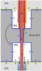
          Apolipoprotein E (apoE) is a major carrier of cholesterol and essential for synaptic plasticity. In brain, it’s expressed by many cells but highly expressed by the choroid plexus and the predominant apolipoprotein in cerebrospinal fluid (CSF). The role of apoE in the CSF is unclear. Recently, the glymphatic system was described as a clearance system whereby CSF and ISF (interstitial fluid) is exchanged via the peri-arterial space and convective flow of ISF clearance is mediated by aquaporin 4 (AQP4), a water channel. We reasoned that this system also serves to distribute essential molecules in CSF into brain. The aim was to establish whether apoE in CSF, secreted by the choroid plexus, is distributed into brain, and whether this distribution pattern was altered by sleep deprivation.

          Methods

          We used fluorescently labeled lipidated apoE isoforms, lenti-apoE3 delivered to the choroid plexus, immunohistochemistry to map apoE brain distribution, immunolabeled cells and proteins in brain, Western blot analysis and ELISA to determine apoE levels and radiolabeled molecules to quantify CSF inflow into brain and brain clearance in mice. Data were statistically analyzed using ANOVA or Student’s t- test.

          Results

          We show that the glymphatic fluid transporting system contributes to the delivery of choroid plexus/CSF-derived human apoE to neurons. CSF-delivered human apoE entered brain via the perivascular space of penetrating arteries and flows radially around arteries, but not veins, in an isoform specific manner (apoE2 > apoE3 > apoE4). Flow of apoE around arteries was facilitated by AQP4, a characteristic feature of the glymphatic system. ApoE3, delivered by lentivirus to the choroid plexus and ependymal layer but not to the parenchymal cells, was present in the CSF, penetrating arteries and neurons. The inflow of CSF, which contains apoE, into brain and its clearance from the interstitium were severely suppressed by sleep deprivation compared to the sleep state.

          Conclusions

          Thus, choroid plexus/CSF provides an additional source of apoE and the glymphatic fluid transporting system delivers it to brain via the periarterial space. By implication, failure in this essential physiological role of the glymphatic fluid flow and ISF clearance may also contribute to apoE isoform-specific disorders in the long term.

          Electronic supplementary material

          The online version of this article (doi:10.1186/s13024-016-0138-8) contains supplementary material, which is available to authorized users.

          Related collections

          Most cited references86

          • Record: found
          • Abstract: found
          • Article: found
          Is Open Access

          A new look at cerebrospinal fluid circulation

          According to the traditional understanding of cerebrospinal fluid (CSF) physiology, the majority of CSF is produced by the choroid plexus, circulates through the ventricles, the cisterns, and the subarachnoid space to be absorbed into the blood by the arachnoid villi. This review surveys key developments leading to the traditional concept. Challenging this concept are novel insights utilizing molecular and cellular biology as well as neuroimaging, which indicate that CSF physiology may be much more complex than previously believed. The CSF circulation comprises not only a directed flow of CSF, but in addition a pulsatile to and fro movement throughout the entire brain with local fluid exchange between blood, interstitial fluid, and CSF. Astrocytes, aquaporins, and other membrane transporters are key elements in brain water and CSF homeostasis. A continuous bidirectional fluid exchange at the blood brain barrier produces flow rates, which exceed the choroidal CSF production rate by far. The CSF circulation around blood vessels penetrating from the subarachnoid space into the Virchow Robin spaces provides both a drainage pathway for the clearance of waste molecules from the brain and a site for the interaction of the systemic immune system with that of the brain. Important physiological functions, for example the regeneration of the brain during sleep, may depend on CSF circulation.
            Bookmark
            • Record: found
            • Abstract: found
            • Article: found
            Is Open Access

            Multiplicity of cerebrospinal fluid functions: New challenges in health and disease

            This review integrates eight aspects of cerebrospinal fluid (CSF) circulatory dynamics: formation rate, pressure, flow, volume, turnover rate, composition, recycling and reabsorption. Novel ways to modulate CSF formation emanate from recent analyses of choroid plexus transcription factors (E2F5), ion transporters (NaHCO3 cotransport), transport enzymes (isoforms of carbonic anhydrase), aquaporin 1 regulation, and plasticity of receptors for fluid-regulating neuropeptides. A greater appreciation of CSF pressure (CSFP) is being generated by fresh insights on peptidergic regulatory servomechanisms, the role of dysfunctional ependyma and circumventricular organs in causing congenital hydrocephalus, and the clinical use of algorithms to delineate CSFP waveforms for diagnostic and prognostic utility. Increasing attention focuses on CSF flow: how it impacts cerebral metabolism and hemodynamics, neural stem cell progression in the subventricular zone, and catabolite/peptide clearance from the CNS. The pathophysiological significance of changes in CSF volume is assessed from the respective viewpoints of hemodynamics (choroid plexus blood flow and pulsatility), hydrodynamics (choroidal hypo- and hypersecretion) and neuroendocrine factors (i.e., coordinated regulation by atrial natriuretic peptide, arginine vasopressin and basic fibroblast growth factor). In aging, normal pressure hydrocephalus and Alzheimer's disease, the expanding CSF space reduces the CSF turnover rate, thus compromising the CSF sink action to clear harmful metabolites (e.g., amyloid) from the CNS. Dwindling CSF dynamics greatly harms the interstitial environment of neurons. Accordingly the altered CSF composition in neurodegenerative diseases and senescence, because of adverse effects on neural processes and cognition, needs more effective clinical management. CSF recycling between subarachnoid space, brain and ventricles promotes interstitial fluid (ISF) convection with both trophic and excretory benefits. Finally, CSF reabsorption via multiple pathways (olfactory and spinal arachnoidal bulk flow) is likely complemented by fluid clearance across capillary walls (aquaporin 4) and arachnoid villi when CSFP and fluid retention are markedly elevated. A model is presented that links CSF and ISF homeostasis to coordinated fluxes of water and solutes at both the blood-CSF and blood-brain transport interfaces. Outline 1 Overview 2 CSF formation 2.1 Transcription factors 2.2 Ion transporters 2.3 Enzymes that modulate transport 2.4 Aquaporins or water channels 2.5 Receptors for neuropeptides 3 CSF pressure 3.1 Servomechanism regulatory hypothesis 3.2 Ontogeny of CSF pressure generation 3.3 Congenital hydrocephalus and periventricular regions 3.4 Brain response to elevated CSF pressure 3.5 Advances in measuring CSF waveforms 4 CSF flow 4.1 CSF flow and brain metabolism 4.2 Flow effects on fetal germinal matrix 4.3 Decreasing CSF flow in aging CNS 4.4 Refinement of non-invasive flow measurements 5 CSF volume 5.1 Hemodynamic factors 5.2 Hydrodynamic factors 5.3 Neuroendocrine factors 6 CSF turnover rate 6.1 Adverse effect of ventriculomegaly 6.2 Attenuated CSF sink action 7 CSF composition 7.1 Kidney-like action of CP-CSF system 7.2 Altered CSF biochemistry in aging and disease 7.3 Importance of clearance transport 7.4 Therapeutic manipulation of composition 8 CSF recycling in relation to ISF dynamics 8.1 CSF exchange with brain interstitium 8.2 Components of ISF movement in brain 8.3 Compromised ISF/CSF dynamics and amyloid retention 9 CSF reabsorption 9.1 Arachnoidal outflow resistance 9.2 Arachnoid villi vs. olfactory drainage routes 9.3 Fluid reabsorption along spinal nerves 9.4 Reabsorption across capillary aquaporin channels 10 Developing translationally effective models for restoring CSF balance 11 Conclusion
              Bookmark
              • Record: found
              • Abstract: found
              • Article: not found

              NG2 cells generate both oligodendrocytes and gray matter astrocytes.

              NG2 glia constitute a fourth major glial cell type in the mammalian central nervous system (CNS) that is distinct from other cell types. Although circumstantial evidence suggests that some NG2 glia differentiate into oligodendrocytes, their in vivo fate has not been directly examined. We have used the bacterial artificial chromosome (BAC) modification technique to generate transgenic mice that express DsRed or Cre specifically in NG2-expressing (NG2+) cells. In NG2DsRedBAC transgenic mice, DsRed was expressed specifically in NG2+ cells throughout the postnatal CNS. When the differentiation potential of NG2+ cells in vitro was examined using DsRed+NG2+ cells purified from perinatal transgenic brains, the majority of the cells either remained as NG2+ cells or differentiated into oligodendrocytes. In addition, DsRed+NG2+ cells also differentiated into astrocytes. The in vivo fate of NG2 glia was examined in mice that were double transgenic for NG2creBAC and the Cre reporter Z/EG. In the double transgenic mice, the Cre reporter EGFP was detected in myelinating oligodendrocytes and in a subpopulation of protoplasmic astrocytes in the gray matter of ventrolateral forebrain but not in fibrous astrocytes of white matter. These observations suggest that NG2+ cells are precursors of oligodendrocytes and some protoplasmic astrocytes in gray matter.
                Bookmark

                Author and article information

                Contributors
                T.rajan.M@gmail.com
                baomanli1226@163.com
                Weiguo_Peng@URMC.Rochester.edu
                Philip.Verghese@gmail.com
                popstone07@gmail.com
                Evan_McConnell@URMC.Rochester.edu
                Abdellatif_Benraiss@URMC.Rochester.edu
                r.tristan.Kasper@gmail.com
                Wei_Song@URMC.Rochester.edu
                Takahiro_Takano@URMC.Rochester.edu
                holtzman@wustl.edu
                Maiken_Nedergaard@URMC.Rochester.edu
                585 273 5377 , Rashid_Deane@urmc.rochester.edu
                Journal
                Mol Neurodegener
                Mol Neurodegener
                Molecular Neurodegeneration
                BioMed Central (London )
                1750-1326
                8 December 2016
                8 December 2016
                2016
                : 11
                : 74
                Affiliations
                [1 ]Center for Translational Neuromedicine, Division of Glial Disease and Therapeutics, Department of Neurosurgery, University of Rochester Medical Center, University of Rochester, Rochester, NY 14642 USA
                [2 ]Department of Neurology, Hope Center for Neurological Disorders, and the Charles F. and Joanne Knight Alzheimer’s Disease Research Center, Washington University School of Medicine, St Louis, MO 63110 USA
                [3 ]Center for Translational Neuromedicine, Division of Cell and Gene Therapy, University of Rochester Medical Center, Rochester, NY 14642 USA
                [4 ]Laboratory of Brain Metabolic Diseases, Institute of Metabolic Disease Research and Drug Development, China Medical University, Shenyang, China
                Article
                138
                10.1186/s13024-016-0138-8
                5146863
                27931262
                009e7397-c62a-4855-b519-25f902264dbd
                © The Author(s). 2016

                Open AccessThis article is distributed under the terms of the Creative Commons Attribution 4.0 International License ( http://creativecommons.org/licenses/by/4.0/), which permits unrestricted use, distribution, and reproduction in any medium, provided you give appropriate credit to the original author(s) and the source, provide a link to the Creative Commons license, and indicate if changes were made. The Creative Commons Public Domain Dedication waiver ( http://creativecommons.org/publicdomain/zero/1.0/) applies to the data made available in this article, unless otherwise stated.

                History
                : 29 April 2016
                : 1 December 2016
                Funding
                Funded by: FundRef http://dx.doi.org/10.13039/100000049, National Institute on Aging;
                Award ID: R21AG050212
                Award Recipient :
                Funded by: FundRef http://dx.doi.org/10.13039/100000065, National Institute of Neurological Disorders and Stroke;
                Award ID: R01 NS078167
                Award ID: R01 NS078304
                Award Recipient :
                Funded by: FundRef http://dx.doi.org/10.13039/100000002, National Institutes of Health;
                Award ID: R01 AG047644
                Award Recipient :
                Categories
                Research Article
                Custom metadata
                © The Author(s) 2016

                Neurosciences
                glymphatic pathways,aqp4,alzheimer’s disease,lymphatic system,brain clearance,sleep/wake

                Comments

                Comment on this article